Journal of Clinical and Investigative Dermatology
Download PDF
Case Report
*Address for Correspondence: Mohammad Ali Alshami, Dermatology Department, Faculty of medicine, and health sciences, Sana’a University, Sana’a, Yemen, Tel: 00967733760082; E-mail: mohammadalshami62@gmail.com
Citation: Alshami MA, Mohana MJ, Alshami AM. Granulosis Rubra Nasi: Case Report. J Clin Investigat Dermatol. 2016;4(1): 2.
Copyright © 2016 Alshami MA, et al. This is an open access article distributed under the Creative Commons Attribution License, which permits unrestricted use, distribution, and reproduction in any medium, provided the original work is properly cited.
Journal of Clinical & Investigative Dermatology | ISSN: 2373-1044 | Volume: 4, Issue: 1
Submission: 26 May, 2016| Accepted: 06 June, 2016 | Published: 10 June, 2016
Granulosis Rubra Nasi: Case Report
Mohammad Ali Alshami1*, Mona Jameel Mohana1 and Ahlam Mohammad Alshami2
- 1Dermatology Department, Faculty of medicine, and health sciences, Sana’a University, Sana’a, Yemen
- 2Faculty of dentistry, Sana’a University, Sana’a, Yemen
*Address for Correspondence: Mohammad Ali Alshami, Dermatology Department, Faculty of medicine, and health sciences, Sana’a University, Sana’a, Yemen, Tel: 00967733760082; E-mail: mohammadalshami62@gmail.com
Citation: Alshami MA, Mohana MJ, Alshami AM. Granulosis Rubra Nasi: Case Report. J Clin Investigat Dermatol. 2016;4(1): 2.
Copyright © 2016 Alshami MA, et al. This is an open access article distributed under the Creative Commons Attribution License, which permits unrestricted use, distribution, and reproduction in any medium, provided the original work is properly cited.
Journal of Clinical & Investigative Dermatology | ISSN: 2373-1044 | Volume: 4, Issue: 1
Submission: 26 May, 2016| Accepted: 06 June, 2016 | Published: 10 June, 2016
Abstract
A 10 years old Yemeni boy presented to the dermatology department with a 4 years history of localized hyperhidrosis, erythema, and telangiectasias over the tip, dorsum, and alae of the nose. On cutaneous examination erythema, covered with beads of sweat, telangiectasias, multiple 2-4 mm transparent cysts, and few erythematous papules were present on the lateral sides and alae of the nose.Based on history and clinical symptoms a diagnosis of granulosis rubra nasi was made.Introduction
Granulosis rubra nasi (GRN) was first described by the German dermatologist Josef Jadassohn in 1901. It is a rare autosomal dominant genodermatosis affecting the eccrine glands of the nose, cheek, and chin. It usually manifests during early childhood, but can manifest in adolescence, and adulthood. It usually starts with localized hyperhidrosis of the tip, dorsum, and alae of nose, yearsbefore other signs appeared. GRN usually disappears spontaneously during puberty. Its diagnosis is based on clinical findings, and biopsyis rarely needed.Case Report
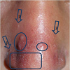
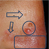
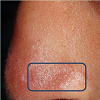
A 10 years old Yemeni boy presented to the dermatology department with a 4 years history of localized hyperhidrosis, erythema, and telangiectasias over the tip, dorsum, and alae of the nose. On cutaneous examination erythema, covered with beads ofsweat, telangiectasias, multiple 2-4 mm transparent cysts, and few erythematous papules were present more on the right side, but also on both alae, and dorsum of the nose (Figures 1a-1c ). The patient was otherwise healthy, and all family members were free of this condition. Based on history and clinical symptoms a diagnosis of granulosis rubra nasi was made.
Discussion
Granulosis rubra nasi (GRN) is an inflammatory condition that involves the eccrine sweat glands of central face and manifests as erythema, localized hyperhidrosis, papules, pustules and vesiclesover central face [1]. GRN was first described by the German dermatologist Josef Jadassohn in 1901 [2]. It is a rare autosomal dominant genodermatosis that usually manifests during early childhood, in patient aged 6 months to 10 years, but can manifest in adolescence, and adulthood. It usually starts with localized hyperhidrosis - seen as small beads of sweat-of the tip, dorsum, and alae of nose, years before other signs appeared. This localized hyperhidrosis seems to be responsible for the secondary changes like erythema and erythematous papules. GRN usually disappeared spontaneously during puberty, but erythema and telangiectasia may persist. Its diagnosis is based on clinical findings, and biopsy is rarely needed [3,4]. The histology shows dilatation of blood vessels, dilated sweat ducts with a discrete mononuclear cell infiltrate surrounding them.The condition must be differentiated from other similar conditions like, acne, rosacea, perioral dermatosis, milia, miliaria, lupus pernio, and lupus erythematosus. In acne there are comedones, papules and pustules distributed symmetrically over the cheeks, forehead, andchin, but there is no localized hyperhidrosis, and no erythema, or telangiectasias. In rosacea there is erythema of the cheeks and nose along with telangiectasias but no hyperhidrosis, in addition to the elder age of rosacea patients. In the case of periorifical dermatitis there involving the perioral and periocular area, leaving a characteristic small free zone around the mouth but no hyperhidrosis. In milia there are longstanding multiple white firm micropapules over both cheeks, but it lacks erythema, and hyperhidrosis. Miliaria crystallina may simulate the beads of sweat but the vesicles cannot be wiped, appear on areas occluded with clothes, and last few days only. Lupus pernio, or chilblain lupus, consists of red or dusky purple papules and plaques not only on the nose, but also on the toes, fingers, and sometimes, elbows, knees and lower legs. The lesions are exacerbated by cold, particularly moist cold climates. Lupus pernio patients have no hyperhidrosis. The bilateral malar erythema “butterfly rash” of acute cutaneous lupus erythematosus (ACLE) is typical, and cannot be confused with GRN. This tends to be transient, follow sun exposure, and resolve without scarring (but sometimes with dyspigmentation). The condition is associated with anti-dsDNA antibodies, lacks hyperhidrosis, and is not confined to the nose. Treatment of granulosis rubra nasi has been described in the literature with topical indomethacin, oral corticosteroids, tetracycline, cryotherapy, and even X-rays [5]. However, reassurance is more important. Grazziotin et al. have recently reported a long-term remission in a patient with granulosis rubra nasi by using botulinum toxin A [6].
A few years ago Piyush Kumar et al. reported the successful use of tacrolimus cream [7].
To conclude GRN is not rare as it seems, but is usually overlooked. An increased awareness of the dermatologist about this condition will lead to early diagnosis. Treatment is not usually needed and reassurance of the patient is enough, as it is self-limited nd disappears during puberty.
References
- Mendoza JP, Saldana LS, Yokota AR, Sialer MC, Anduaga ES (2003) Nasi rubra granulosis. Dermatol Peru 13: 125-127.
- Jadasson J (1901) Ueber eine eigenartige Erkrankung der Nasenhaut bei Kindern (“Granulosis rubra nasi”). Archiv Dermatol und Syph 58: 145-158.
- Sargunam C, Thomas J, Ahmed NA (2013) Granulosis rubra nasi. Indian Dermatol Online J 4: 208-209.
- Sonthalia S, Singal A, Sharma R (2012) Hyperhidrosis, vesicles, and papules over the nose: granulosis rubra nasi. Indian J Dermatol Venereol Leprol 78: 97-98.
- Hellier FF (1937) Granulosis rubra nasi in a mother and daughter. Br Med J 2: 1068.
- Grazziotin TC, Buffon RB, da Silva Manzoni AP, Libis AS, Weber MB (2009) Treatment of granulosis rubra nasi with botulinum toxin type A. Dermatol Surg 35: 1298-1299.
- Kumar P, Gosai A, Mondal AK, Lal NR, Gharami RC (2012) Granulosis rubra nasi: a rare condition treated successfully with topical tacrolimus. Dermatol Reports 4: e5.


