
Journal of Clinical and Investigative Dermatology
Download PDF
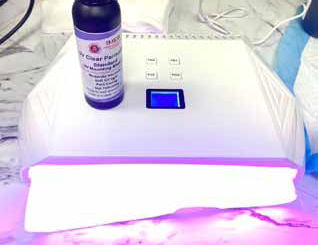 Figure 1: UV Clear PermaSlip Mounting Medium and a UV light trialed as a new clearing agent in our clinic.
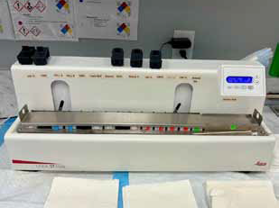 Figure 2: Leica ST4020 linear slide stainer and stains used in our clinic.
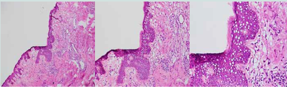 Figure 3: Tissue sample using UV mouting medium and UV light under the miscroscope at (from left to right) 10X, 20X, and 40X.
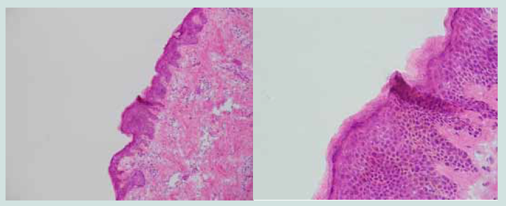 Figure 4: Same tissue sample using xylene substitiute under the miscroscope at 10X (left) and 40X (right).
Research Article
Trial of Ultraviolet Clear PermaSlip Mounting Medium and Ultraviolet Light as New Clearing Agent in Mohs Micrographic Surgery Tissue Processing
Kichena S1, Aldrete J2 and Tolkachjov SN3-6*
1Paul L. Foster School of Medicine, Texas Tech University Health Sciences Center, El Paso, TX
2Department of Dermatology, Texas Tech University Health Sciences Center, Lubbock, Texas
3Department of Dermatology, Baylor University Medical Center, Dallas, Texas
4Epiphany Dermatology, Dallas, Texas
5Department of Dermatology, University of Texas at Southwestern, Dallas, Texas
6Texas A&M University College of Medicine, Dallas, Texas
*Address for Correspondence: Stanislav N. Tolkachjov, Department of Dermatology, University
of Texas at Southwestern, Dallas, Texas USA. E mail Id: Stan. tolkachjov@gmail.com
Submission:22 July, 2024
Accepted:15 August, 2024
Published:20 August, 2024
Copyright: © 2024 Kichena S, et al. This is an open access article
distributed under the Creative Commons Attri-bution License,
which permits unrestricted use, distribution, and reproduction in
any medium, provided the original work is properly cited.
Keywords:Clearing Agent; UV Mounting Medium; UV Light; Xylene;
Xylene Substitute
Abstract
Xylene and xylene substitutes are commonly used as clearing
agents when processing tissue samples during Mohs micrographic
surgery (MMS). However, there are several concerns with these solvents.
Xylene is expensive, malodorous, and can be toxic depending on
exposure levels. While xylene substitutes are less toxic, some of these
are more expensive than xylene itself or do not biodegrade easily and
require specific waste disposal. Here, we discuss an alternative using
Ultraviolet (UV) Clear PermaSlip Mounting Medium and a UV light. This
technique is advantageous in that it is non-toxic, has no odor, and does
not have the significant costs of purchase and disposal that xylene and
xylene substitute typically have. However, we found this technique
can add up to 2 minutes to the process, and more importantly, may
reduce the quality of the slide. Upon slide review with this method,
we found more intracellular lacunae artifact with keratinocytes in the
epidermis compared to the same tissue on consecutive cuts where
traditional xylene substitute was used. Future studies could look at ways
to minimize lacunae with this method. Ultimately, we must weigh the
advantages and disadvantages of using a UV mounting medium and
UV light and utilize our different options to provide the best quality
patient care
Abbreviations
MMS: Mohs Micrographic Surgery; UV: Ultraviolet
Introduction
MMS is a specialized form of skin cancer surgery resulting in a
nearly 100% cure rate while minimizing lost tissue and is particularly
useful in cosmetically sensitive areas [1]. The surgeon first removes
a thin layer of skin from the skin cancer. The tissue is then frozen,
cut into thin slices, stained, and evaluated under a microscope for
circumferential and deep tissue margin clearance of the skin cancer.
If the margins are not clear, the process is repeated until they are
clear [2]. During this tissue processing, speed and efficiency are
pertinent in providing quality care. The histologicfrozen-tissue stains
commonly used are generally based on preference and training,
but three commonly used stains are toluidine blue, thionine, and
hematoxylin &eosin [3]. Today, many Mohs clinics use automated
slide stainers. Afterwards, a clearing agent is used to make biological
tissues transparent while preserving tissue structure, allowing for
visualization of the tissue under a microscope [4].
Currently, there are three main categories of tissue clearing
techniques: solvents, hyperhydration, and hydrogel embedding
techniques [4]. However, hyperhydration and hydrogel
embedding technique are very time consuming and can take days.
One such solvent used as a clearing agent is xylene. Xylene is an
aromatic hydrocarbon commonly used during tissue processing in
MMS. Advantages of xylene usage include that it is biodegradable,
noncorrosive, nonflammable, soluble in alcohol and mounting media,
reasonably fast drying, and doesn’t leave a residue. However, xylene is
also expensive, malodorous, and can be toxic depending on exposure
levels. Some literature even recommends the use of a fume hood or
some local exhaust ventilation in addition to personal protective
equipment to reduce toxicity [5] .MMS labs now commonly use
solvents such as Naphthenic solvent and d-limonenes chemicals that
can be used a xylene substitute, but some of these are more expensive
than xylene itself or do not biodegrade easily and require specific
waste disposal.
Materials and Methods
As a result, in our clinic, we trialed the use of UV Clear PermaSlip
Mounting Medium and a UV light as a new clearing agent [Figure 1].
Using the Leica ST4020 linear slide stainer, we utilized a combination
of alcohol, H2O, Gill’s Hematoxylin I, Gill’s Hematoxylin II, Toluidine
blue, and Eosin Y during the staining process [Figure 2]. In order
to use this mounting medium, the slide must first dry after going
through the staining process. Then a small amount of the mounting
medium is applied to the slide and cover slipped. Finally, the slide
must sit under a UV light for approximately 30 seconds. This whole
process can take up to 2 minutes.
Results
We found that this technique is advantageous in that it is nontoxic,
has no odor, and does not have the significant costs of purchase
and disposal that xylene and xylene substitute typically have. However,
we found this technique can add up to 2 minutes to the process, and
more importantly, may reduce the quality of the slide. Upon slide
review with this method, we found more intracellular lacunae artifact
with keratinocytes in the epidermis [Figure 3] compared to the same
tissue on consecutive cuts where traditional xylene substitute was
used [Figure 4]. These intracellular lacunae are likely the result of
focal evaporation and are important as melanocytic lesions, some
inflammatory conditions, and extramammary Paget’s disease may be
confused with this artifact.
We present another viable clearing method that can be utilized
in MMS tissue processing. It is important to weigh the advantages
and disadvantages of using a UV mounting medium and UV light in
comparison to xylene or xylene substitutes. For example, in a clinic
trying to cut the use of xylene substitutes or cost, UV mounting
medium in conjunction with a UV light may be beneficial. Future
studies could look at ways to minimize lacunaewith this method.
Drying the slides for a longer duration, using a different drying
method, and/or changing the amount of time spent under UV light
may reduce this artifact. Ultimately, we must utilize our different
options toprovide the best quality patient care.
Acknowledgements
Dr. Tolkachjov is an investigator and speaker for Bioventus and
CASTLE and advisory board member for Illumisonics. Other authors
have no relevant conflicts of interest to declare and did not receive
any grants or funding for this project.
References
Citation
Kichena S, Aldrete J, Tolkachjov SN. Trial of Ultraviolet Clear PermaSlip Mounting Medium and Ultraviolet Light as New Clearing Agent in Mohs Micrographic Surgery Tissue Processing. J Clin Investigat Dermatol. 2024;12(1): 1