Journal of Gene Therapy
Comparative Analysis of H1N1 Avian Influenza Virus by Multiple Sequence Alignment and Support Vector Machine
Yufei Liu, Libin Zhang* and Yanhong Zhou*
- Department of Biomedical Engineering, School of Life Science and Technology, Huazhong University of Science and Technology, Wuhan, Hubei 430074, China
*Address for Correspondence: Libin Zhang, Department of Biomedical Engineering, School of Life Science and Technology, Huazhong University of Science and Technology, Wuhan, Hubei 430074, China; E-mail: libinzhang@hust.edu.cn
Yanhong Zhou, Department of Biomedical Engineering, School of Life Science and Technology, Huazhong University of Science and Technology, Wuhan, Hubei 430074, China; E-mail: E-mail: yhzhou@hust.edu.cn
Citation: Liu Y, Zhang L, Zhou Y. Comparative Analysis of H1N1 Avian Influenza Virus by Multiple Sequence Alignment and Support Vector Machine. J Gene Ther 2014; 2(1): 4.
Copyright © 2014 Liu Y, et al. This is an open access article distributed under the Creative Commons Attribution License, which permits unrestricted use, distribution, and reproduction in any medium, provided the original work is properly cited.
Journal of Gene Therapy | ISSN: 2381-3326 | Volume: 1, Issue: 1
Submission: 11 September 2013 | Accepted: 03 January 2014 | Published: 10 January 2014
Reviewed & Approved by: Dr. Yujing Li, Senior Scientist, Emory University School of Medicine, USA
Abstract
The H1N1 is a subtype of avian influenza virus (AIV) that is able to break the host barrier to seriously endanger human health. Investigating the molecular mechanisms of AIV interspecies transmission is important for preventing the influenza epidemics. In this study, we used bioinformatics approaches to identify factors that may cause the avian-to-human transmission in hemagglutinin (HA) sequences of H1N1. First, the multiple sequence alignment analysis reveals 10 signature regions of HA that are highly conserved in intra-species, but largely divergent between interspecies. Then, the avian-to-human transformation was modeled as a binary classification problem in a machine learning (ML) context. A computational prediction model was developed to predict the avian-to-human transmission of H1N1 with advanced ML techniques by characterizing amino acid residues in these signatures regions. The evaluation results suggested that these amino acid residues have a discrimination ability to distinguish H1N1 strains isolated from human to those from avian. The proposed bioinformatic framework would be helpful for further understanding the transmission mechanisms of H1N1 and other AIV viruses.
Keywords
Avian influenza virus; Bioinformatics; H1N1; Hemagglutinin; Machine learning; Support vector machine; Sequence alignmentIntroduction
Influenza is a paramount epidemic in the world because of the continuing evolution of virus via antigenic drift and genetic shift [1]. The avian influenza virus is infectious for birds, pigs, horses and human and lead to the lesions of avian body or respiratory tract, which does great harm to the breeding of poultry such as chickens, turkeys and ducks etc [2-6]. Therefore, the study of avian influenza virus not only has great significance to the poultry industry but also to human health.
H1N1 is the subtype of influenza A virus (AIV) that can break the host barrier to seriously endanger human health, exemplified by the 2009 swine-origin H1N1 influenza A epidemic [7]. Although the origins and evolutionary of human-isolated H1N1 virus can be easily inferred from the phylogenic analysis, the determinants of cross-species transmission are still not fully understood [8,9]. The H1N1 genome codes six internal proteins (NP, M1, M2, PB1, PB2 and PA), two non-structural proteins (NS1 and NS2), and two coat proteins (HA and NA) [10]. Among these ten proteins, HA (hegagglutinin) has been demonstrated to be particularly important for virus infection against the host, by mediating the attachment of the virus to the host cell surface and the entry of viral RNA into the host cell [11,12]. Therefore, the properties of the HA protein in H1N1 virus are very worthwhile to be studied, which will provide a clue to better understand the infection mechanism of influenza viruses and monitor the interspecies transmission of influenza virus.
In this study, we used bioinformatic approaches to comparatively analyze the HA protein sequences of H1N1 viruses isolated from avian and human hosts, and identified several signature regions in which amino acid segments specifically are conserved in avian- and human- isolated H1N1 viruses. Further statistical analysis revealed that there are different patterns of amino acid content in the HA sequences of H1N1 viruses isolated before and after the 2009 H1N1 pandemic. We also applied machine learning techniques to effectively distinguishhuman-isolated H1N1 from avian-isolated H1N1.
Methods
Multiple sequence alignment
In bioinformatics, multiple sequence alignment is used for arranging the sequences of DNA, RNA, or protein to identify similar regions that may conclude the important conclusions of function, structure, or evolution of species [13]. Multiple sequence alignment aligned all of the sequences to a unified format for analyzing the functional or structural regions in samples. Multiple sequence alignment is also the necessary steps to construct phylogenetic trees for aiding in identifying evolutionary relationships [14,15]. In this study, the multiple sequence alignment was performed with online alignment software integrated into the NCBI Influenza virus resource (http://www.ncbi.nlm.nih.gov/genomes/FLU/FLU.html).
Support vector machine
The Support Vector Machine (SVM) is a well-formulated machine learning technology first proposed by Corinna Cortes and Vapnik in 1995 [16]. The SVM mapped the features in the original space into a high-dimensional feature space with a kernel function to perform the classification problems. Due to the advantages of solving small samples, nonlinear and high-dimensional pattern recognition, it has been applied for various classification and prediction problems, including the classification of translation initiation starts [17], protein subcellular localization [18] and protein function [19]. In this study, we performed the SVM algorithm by considering the amino acid content in signature regions through implementing the ‘e1071’ package with the default parameters in R programming language (http://cran.r-project.org).
Results
Signature regions identified in HA genes of H1N1 by multiple sequence alignment analysis
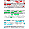
All HA sequences of the H1N1 avian influenza virus isolated from human (3377) and avian (127) analyzed in this study were downloaded from the Influenza Virus Resource of NCBI. Examining the host specificity of amino acid (AA) residues is helpful to identify important regions that may play roles in the cross-transmission of H1N1 in the HA sequence. The AA residues in a successive positions were consider to be host specific if they were highly conserved in the same species of the host, but evidently divergent between two species. It should be noted that this definition of host specificity of AA residues can also be applied on other virus proteins and at the structure level. We found that several regions (denoted as signature regions) in HA sequences were host specific (Table 1). At the regions of 489~490 and 144~152, the most frequency of AA residues in avian-isolated H1N1 viruses were “DDE” (94.6%) and “ETTKGVTAA” (93%), respectively. While in human-isolated H1N1 viruses, the corresponding frequency were about 61%. This result indicated that amino acids of this signature region in human-isolated H1N1 viruses evolved more rapidly than those in avian-isolated H1N1 viruses. We also observed that at the region of 9~16, the frequencies of “FCTFTVLK” residues were comparable between H1N1 isolated from avian and human hosts (65% vs 63%). Further statistical analysis revealed that nearly 60% contained all the identified amino acid residues (Figure 1A), red amino acids, allowed for 2 amino acids mismatch) among 3377 H1N1 strains in human. On the other hand, the majority of H1N1 strains in avian (total number, 127) were found to contain all the identified amino acids (Figure 1C, blue amino acids, allowed for 2 amino acids mismatch). Finally, NA protein was selected for same analysis as HA protein. We picked out a total of 904 H1N1 strains from human to perform multiple protein sequence alignment and the amino acid frequencies in 470 sites were counted. As shown in Supplementary Table 1, the result showed that the majority of amino acid frequencies in 470 sites are more than 80% except for sites 241, 248 and 369. Moreover, the number of the sites in which the frequencies of amino acids are more than 85% is 451, which indicated that the multiple sequence alignment analysis of NA protein displayed a single pattern in human-isolated H1N1 viruses.
Figure 1: Pattern classification of the H1N1 avian influenza virus. (A). Pattern I of the H1N1 avian influenza virus in human (containing all the amino acids marked in red, allow for 2 amino acids mismatch). (B). Pattern II of the H1N1 avian influenza virus in human (containing all the amino acids marked in green, allow for 2 amino acids mismatch). (C).The major pattern of H1N1 avian influenza virus in avian (containing all the amino acids marked in blue, allow for 2 amino acids mismatch).
Support vector machine verification
By drawing the identified signature regions as the characteristics and setting H1N1 strains in human as the negative samples and H1N1 strains in avian as the positive samples, we built up a classification mode between avian-isolated and human-isolated H1N1 with SVM. Although the classification of AIV from avian and human hostshas been recently investigated [20,21], we further performed the classification problems on H1N1 strains isolated in different years. As shown in Table 2, the result revealed that the HA fragments we picked up can be used to identify the differences between isolated- and human-isolated H1N1 viruses, and suggested that SVM is an effective classifier for distinguishing the H1N1 influenza viruses isolated from avian and human hosts. In addition the SVM analysis among different H1N1 species in human was shown in Table 3. On the basis of Table 3, the avian influenza virus in human was obviously different every year. This result not only indicated influenza viruses evolved very fast, but also confirmed that H1N1 influenza virus with different species and same species at different times were both distinguished by HA fragments.
Discussion
In this study, we performed the multiple sequence alignment analysis to identify several signature regions of HA in which the frequencies of amino acid fragments in human-isolated H1N1 were different from those in avian-isolated H1N1. Using machine learning technique, the amino acids in these signature regions helped us to build prediction model for classifying the avian- and human-isolated H1N1 viruses.
The multiple sequence alignment analysis also helped us to identify three patterns of amino acid content of HA protein in human- and avian-isolated H1N1 viruses. As shown in Figure 1A, statistical analysis demonstrated that nearly 60% contained all the amino acids (marked in red, allow for 2 amino acids mismatch) among 3377 H1N1 strains in human. We described these human H1N1 strains as pattern I H1N1 virus in human (Figure 1A). On the other hand, nearly 40% H1N1 strains in human contained all the mino acids (marked in green, allow for 2 amino acids mismatch) and were described as pattern II H1N1 virus in human (Figure 1B). Also, statistical analysis demonstrated that majority of H1N1 strains in avian (total number, 127) contained all the amino acids (marked in blue, allow for 2 amino acids mismatch) and were described as major H1N1 pattern in avian (Figure 1C). Most of pattern I H1N1 influenza viruses in human appeared recently after the 2009 AIV pandemic. Moreover, more than 90% of human H1N1 viruses ppeared in Europe and Asia after 2009 were classified into pattern I. On the contrary, Most of pattern II H1N1 influenza viruses in human appeared before 2009 and the number of viruses decreased very quickly after 2009. Further investigation of these patterns would be helpful for understanding the evolution of H1N1, and forecastingthe potential AIV pandemic. Different from HA protein, the multiple sequence alignment analysis of NA protein (Supplementary Table 1) did not display the two patterns (pattern I and II, please discussion) appearing in HA protein analysis, which suggested that NA is more conserved than HA and HA plays a more important role in the interspecies transmission process of avian influenza virus.
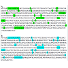
In the meanwhile, H7N9 sequences deposited in NCBI database including 9 strains isolated from human and 39 strains isolated from poultry were downloaded for multiple sequence alignment analysis. The result showed that the strains from poultry also displayed two patterns (pattern I and pattern II). As shown in Figure 2, pattern I has different conserved amino acid sites from pattern II and the pattern I strains have high sequence similarity with strains from human. Furthermore, the similarity of amino acids between pattern I strains and strains from human added up to 89.8%. Therefore, we speculated that pattern I H7N9 strains from poultry are easier to infect human and need to be monitored. Although the number of collected H7N9 strains is limited, the obtained result can be used as a reference for further study. Collectively, the bioinformatics framework applied in this study will facilitate in-depth knowledge discovery of transmission mechanisms from the protein sequences of H1N1 viruses.
Figure 2: Pattern classification of the H7N9 avian influenza virus. (A). Pattern I of the H7N9 avian influenza virus in avian (containing all the amino acids marked in green, allow for 2 amino acids mismatch). (B). Pattern II of H7N9avian influenza virus in avian (containing all the amino acids marked in blue, allow for 2 amino acids mismatch).
References
- Subbarao K, Joseph T (2007) Scientific barriers to developing vaccines against avian influenza viruses. Nat Rev Immunol 7: 267-278.
- Webster RG, Bean WJ, Gorlnan OT, Chambers TM, Kawaoka Y (1992) Evolution and ecology of influenza A viruses. Microbiol Rev 56: 152-179.
- Sorek R, Kunin V, Hugenholtz P (2008) CRISPR-a widespread system that provides acquired resistance against phages in bacteria and archaea. Nat Rev Microbiol 6:181-186.
- Garneau JE, Dupuis MÈ, Villion M, Romero DA, Barrangou R, et al. (2010) The CRISPR/Cas bacterial immune system cleaves bacteriophage and plasmid DNA. Nature 468: 67-71.
- Wilson RC, Doudna JA (2013) Molecular mechanisms of RNA interference. Annu Rev Biophys 42: 217-39.
- Brantl S (2002) Antisense-RNA regulation and RNA interference. Biochim Biophys Acta 1575:15-25.
- Waters LS, Storz G (2009) Regulatory RNAs in bacteria. Cell 136: 615-628.
- Gerdes K, Wagner EGH (2007) RNA antitoxins. Curr Opin Microbiol 10:117-24.
- Dornenburg JE, Devita AM, Palumbo MJ, Wade JT (2010) Widespread antisense transcription in Escherichia coli. MBio 1: e00024-10. 1 5
- Sharma CM, Hoffmann S, Darfeuille F, Reignier J, Findeiss S, et al. (2010) The primary transcriptome of the major human pathogen Helicobacter pylori. Nature 464: 250-255.
- Han Y, Lin YB, An W, Xu J, Yang HC, et al. (2008) Orientation-dependent regulation of integrated HIV-1 expression by host gene transcriptional read through. Cell Host Microbe 4:134-46.
- Liu JM, Livny J, Lawrence MS, Kimball MD, Waldor MK, et al. (2009) Experimental discovery of sRNAs in Vibrio cholerae by direct cloning, 5S/tRNA depletion and parallel sequencing. Nucleic Acids Res 37: e46.
- Toledo-Arana A, Dussurget O, Nikitas G, Sesto N, Guet-Revillet H, et al. (2009) The Listeria transcriptional landscape from saprophytism to virulence. Nature 459:950-956.
- Güell M, van Noort V, Yus E, Chen WH, Leigh-Bell J, et al. (2009) Transcriptome Complexity in a Genome-Reduced Bacterium. Science326:1268-1271.
- Georg J, Voss B, Scholz I, Mitschke J, Wilde A, et al. (2009) Evidence for a major role of antisense RNAs in cyanobacterial gene regulation. Mol Syst Biol 5:305.
- Katayama S, Tomaru Y, Kasukawa T, Waki K, Nakanishi M, et al. (2005) Antisense transcription in the mammalian transcriptome. Science 309: 1564-1566.
- Yelin R, Dahary D, Sorek R, Levanon EY, Goldstein O, et al. (2003) Widespread occurrence of antisense transcription in the human genome. Nat Biotechnol 21: 379-86.
- Misra S, Crosby MA, Mungall CJ, Matthews BB, Campbell KS, et al. (2002) Annotation of the Drosophila melanogaster euchromatic genome: asystematic review. Genome Biol 3: 1-22.
- Yamada K, Lim J, Dale JM, Chen H, Shinn P, et al. (2003) Empirical analysis of transcriptional activity in the Arabidopsis genome. Science 302: 842-846.
- Berteaux N, Aptel N, Cathala G, Genton C, Coll J, et al. (2008) A novel H19 antisense RNA overexpressed in breast cancer contributes to paternal IGF2 expression. Mol Cell Biol 28: 6731-45.
- Monti L, Cinquetti R, Guffanti A, Nicassio F, Cremona M, et al. (2009) In silico prediction and experimental validation of natural antisense transcripts in two cancer-associated regions of human chromosome 6. Int J Oncol 34: 1099-108.
- Marshall L, White RJ (2008) Non-coding RNA production by RNA polymerase III is implicated in cancer. Nat Rev Cancer 8: 911-914.
- Coiras M, López-Huertas MR, Pérez-Olmeda M, Alcamí J (2009) Understanding HIV-1 latency provides clues for the eradication of long-term reservoirs. Nat Rev Microbiol 7:798-812.
- Kröger C, Dillon SC, Cameron AD, Papenfort K, Sivasankaran SK, et al. (2012) The transcriptional landscape and small RNAs of Salmonella enterica serovar Typhimurium. Proc Natl Acad Sci U S A 109: E1277-86.
- Mitschke J, Georg J, Scholz I, Sharma CM, Dienst D, et al. (2011) An experimentally anchored map of transcriptional start sites in the model cyanobacterium Synechocystis sp. PCC6803. Proc Natl Acad Sci U S A 108: 2124-2129.
- Nicolas P, Mäder U, Dervyn E, Rochat T, Leduc A, et al. (2012) Condition-Dependent Transcriptome Reveals High-Level Regulatory. Science 335: 1103-1106.
- Albrecht M, Sharma CM, Reinhardt R, Vogel J, Rudel T (2010) Deep sequencing-based discovery of the Chlamydia trachomatis transcriptome.Nucleic Acids Res 38: 868-877.
- Wurtzel O, Yoder-Himes DR, Han K, Dandekar AA, Edelheit S, et al. (2012) The single-nucleotide resolution transcriptome of Pseudomonas aeruginosa grown in body temperature. PLoS Pathog 8: e1002945.
- Wurtzel O, Yoder-Himes DR, Han K, Dandekar AA, Edelheit S, et al. (2010) Transcriptome analysis of Pseudomonas syringae identifies new genes, noncoding RNAs, and antisense activity. J Bacteriol 192: 2359-72.
- Beaume M, Hernandez D, Farinelli L, Deluen C, Linder P, et al. (2010) Cartography of methicillin-resistant S. aureus transcripts: detection,orientation and temporal expression during growth phase and stressconditions. PLoS One 5: e10725.
- Schlüter JP, Reinkensmeier J, Daschkey S, Evguenieva-Hackenberg E, Janssen S, et al. (2010) A genome-wide survey of sRNAs in the symbiotic nitrogen-fixing alpha-proteobacterium Sinorhizobium meliloti. BMC Genomics 11: 245.
- Chatterjee A, Johnson CM, Shu CC, Kaznessis YN, Ramkrishna D, et al. (2011) Convergent transcription confers a bistable switch in Enterococcus faecalis conjugation. Proc Natl Acad Sci U S A 108:9721-9726.
- Hongay CF, Grisafi PL, Galitski T, Fink GR (2006) Antisense transcription controls cell fate in Saccharomyces cerevisiae. Cell 127:735-45.
- Georg J, Hess WR (2011) cis-antisense RNA, another level of gene regulation in bacteria. Microbiol Mol Biol Rev 75: 286-300.
- Shearwin KE, Callen BP, Egan JB (2005) Transcriptional interference--a crash course. Trends Genet 21: 339-345.
- Thomason MK, Storz G (2010) Bacterial antisense RNAs: how many are there, and what are they doing? Annu Rev Genet 44:167-188.
- Callen BP, Shearwin KE, Egan JB (2004) Transcriptional Interference between Convergent Promoters Caused by Elongation over the Promoter.Mol Cell 14: 647-656.
- Palmer AC, Ahlgren-Berg A, Egan JB, Dodd IB, Shearwin KE (2009) Potent transcriptional interference by pausing of RNA polymerases over a downstream promoter. Mol Cell 34: 545-55.
- Ward DF, Murray NE (1979) Convergent Transcription in Bacteriophage lambda: Interference with Gene Expression. J Mol 133: 249-266.
- Greger IH, Aranda A, Proudfoot N (2000) Balancing transcriptional interference and initiation on the GAL7 promoter of Saccharomyces cerevisiae. PNAS 97: 8415-8420.
- Gullerova M, Proudfoot NJ (2008) Cohesin complex promotes transcriptional termination between convergent genes in S. pombe. Cell 132: 983-95.
- Franch T, Petersen M, Wagner EG, Jacobsen JP, Gerdes K (1999) Antisense RNA regulation in prokaryotes: rapid RNA/RNA interaction facilitated by a general U-turn loop structure. J Mol Biol 294: 1115-1125.
- Bennett CF, Swayze EE (2010) RNA targeting therapeutics: molecular mechanisms of antisense oligonucleotides as a therapeutic platform. Annu Rev Pharmacol Toxicol 50: 259-293.
- Johnson E, Srivastava R (2012) Volatility in mRNA secondary structure as a design principle for antisense. Nucleic Acids Res 41: 1-10.
- Yamaguchi Y, Park J-H, Inouye M (2011) Toxin-antitoxin systems in bacteria and archaea. Annu Rev Genet 45: 61-79.
- Brantl S (2007) Regulatory mechanisms employed by cis-encoded antisense RNAs. Curr Opin Microbiol 10: 102-9.
- Sneppen K, Dodd IB, Shearwin KE, Palmer AC, Schubert RA, et al. (2005) A mathematical model for transcriptional interference by RNA polymerase traffic in Escherichia coli. J Mol Biol 346: 399-409.
- Palmer AC, Egan JB, Shearwin KE (2011) Transcriptional interference by RNA polymerase pausing and dislodgement of transcription factors.Transcription 2: 9-14.
- Lee EJ, Groisman EA (2010) An antisense RNA that governs the expression kinetics of a multifunctional virulence gene. Mol Microbiol 76: 1020-1033.
- Thisted T, Gerdes K (1997) Mechanism of Post-segregational Killing of Plasmid Rl by the hok/sok System Sok Antisense RNA Regulates hok Gene Expression Indirectly Through the Overlapping mok Gene. J Mol Biol: 41-54.
- Hernández JA, Muro-Pastor AM, Flores E, Bes MT, Peleato ML, et al. (2006) Identification of a furA cis-antisense RNA in the cyanobacterium Anabaena sp. PCC 7120. J Mol Biol 355: 325-34.
- Liao SM, Wu TH, Chiang CH, Susskind MM, McClure WR (1987) Control of gene expression in bacteriophage P22 by a small antisense RNA. I.Characterization in vitro of the Psar promoter and the sar RNA transcript. Genes Dev 1: 197-203.
- Kawano M, Aravind L, Storz G (2007) An antisense RNA controls synthesis of an SOS-induced toxin evolved from an antitoxin. Mol Microbiol 64: 738-54.
- Krinke L, Wulff DL (1987) OOP RNA, produced from multicopy plasmids, inhibits lambda cII gene expression through an RNase III-dependentmechanism. Genes Dev 1: 1005-1013.
- Stazic D, Lindell D, Steglich C (2011) Antisense RNA protects mRNA from RNase E degradation by RNA-RNA duplex formation during phage infection. Nucleic Acids Res 39: 4890-4899.
- Montange RK, Batey RT (2008) Riboswitches: emerging themes in RNA structure and function. Annu Rev Biophys 37: 117-133.
- Wan Y, Kertesz M, Spitale RC, Segal E, Chang HY (2011) Understanding the transcriptome through RNA structure. Nat Rev Genet 12: 641-655.
- Brantl S (2002) Antisense-RNA regulation and RNA interference. Biochim Biophys Acta - Gene Struct Expr 1575: 15-25.
- Arraiano CM, Andrade JM, Domingues S, Guinote IB, Malecki M, et al. (2010) The critical role of RNA processing and degradation in the control of gene expression. FEMS Microbiol Rev 34: 883-923.
- Johnson CM, Manias DA, Haemig HA, Shokeen S, Weaver KE, et al. (2010) Direct evidence for control of the pheromone-inducible prgQ operon of Enterococcus faecalis plasmid pCF10 by a counter transcript-driven attenuation mechanism. J Bacteriol 192: 1634-42.
- Sayed N, Jousselin A, Felden B (2012) A cis-antisense RNA acts in trans in Staphylococcus aureus to control translation of a human cytolytic peptide. Nat Struct Mol Biol 19: 105-112.
- Stork M, Di Lorenzo M, Welch TJ, Crosa JH (2007) Transcription termination within the iron transport-biosynthesis operon of Vibrio anguillarum requires an antisense RNA. J Bacteriol 189: 3479-3488.
- Opdyke JA, Fozo EM, Hemm MR, Storz G (2011) RNase III participates in GadY-dependent cleavage of the gadX-gadW mRNA. J Mol Biol 406: 29-43.
- Dühring U, Axmann IM, Hess WR, Wilde A (2006) An internal antisense RNA regulates expression of the photosynthesis gene isiA. Proc Natl Acad Sci U S A 103: 7054-7058.
- Johnson CM, Haemig HH, Chatterjee A, Wei-Shou H, Weaver KE, et al. (2011) RNA-Mediated Reciprocal Regulation between Two Bacterial Operons is RNase III Dependent. MBio 2: e00189-11.
- Prescott EM, Proudfoot NJ (2002) Transcriptional collision between convergent genes in budding yeast. Proc Natl Acad Sci 99: 8796-8801.
- Crampton N, Bonass WA, Kirkham J, Rivetti C, Thomson NH (2006) Collision events between RNA polymerases in convergent transcription studied by atomic force microscopy. Nucleic Acids Res 34: 5416-5425.
- Shu CC, Chatterjee A, Dunny G, Hu WS, Ramkrishna D (2011) Bistability versus bimodal distributions in gene regulatory processes from population balance. PLoS Comput Biol 7: e1002140.
- Chatterjee A, Drews L, Mehra S, Takano E, Kaznessis YN, et al. (2011) Convergent transcription in the butyrolactone regulon in Streptomycescoelicolor confers a bistable genetic switch for antibiotic biosynthesis. PLoS One 6: e21974.
- Palmer AC, Ahlgren-Berg A, Egan JB, Dodd IB, Shearwin KE (2009) Potent transcriptional interference by pausing of RNA polymerases over a downstream promoter. Mol Cell 34: 545-555.
- Bendtsen KM, Erdossy J, Csiszovszki Z, Svenningsen SL, Sneppen K, et al. (2011) Direct and indirect effects in the regulation of overlapping promoters. Nucleic Acids Res 39: 6879-6885.
- Mason E, Henderson MW, Scheller EV, Byrd MS, Cotter PA (2013) Evidence for phenotypic bistability resulting from transcriptional interference of bvgAS in Bordetella bronchiseptica. Mol Microbiol 90: 716-733.
- Stougaard P, Molin S, Nordstrom K (1981) RNAs involved in copy-number control and incompatibility of. Proc Natl Acad Sci 78: 6008-6012.
- Tomizawa J, Itoh T (1981) Plasmid ColE1 incompatibility determined by interaction of RNA I with primer transcript. Proc Natl Acad Sci 78: 6096-6100.
- Hernández JA, Muro-Pastor AM, Flores E, Bes MT, Peleato ML, et al. (2006) Identification of a furA cis-antisense RNA in the cyanobacterium Anabaena sp. PCC 7120. J Mol Biol 355: 325-334.
- Stazic D, Lindell D, Steglich C (2011) Antisense RNA protects mRNA from RNase E degradation by RNA-RNA duplex formation during phage infection. Nucleic Acids Res 39: 4890-4899.
- Toledo-Arana A, Dussurget O, Nikitas G, Sesto N, Guet-Revillet H, et al. (2009) The Listeria transcriptional landscape from saprophytism to virulence. Nature 459: 950-956.
- André G, Even S, Putzer H, Burguière P, Croux C, et al. (2008) S-box and T-box riboswitches and antisense RNA control a sulfur metabolic operon of Clostridium acetobutylicum. Nucleic Acids Res 36: 5955-5969.
- Chatterjee A, Cook LC, Shu CC, Chen Y, Manias DA, et al. (2013) Antagonistic self-sensing and mate-sensing signaling controls antibiotic resistance transfer. Proc Natl Acad Sci U S A 110: 7086-7090.
- Cook L, Chatterjee A, Barnes A, Yarwood J, Hu WS, et al. (2011) Biofilm growth alters regulation of conjugation by a bacterial pheromone. Mol Microbiol 81: 1499-1510.
- Giangrossi M, Prosseda G, Tran CN, Brandi A, Colonna B, et al. (2010) A novel antisense RNA regulates at transcriptional level the virulence gene icsA of Shigella flexneri. Nucleic Acids Res 38: 3362-3375.6.
- Chen J, Sun M, Hurst LD, Carmichael GG, Rowley JD (2005) Genomewideanalysis of coordinate expression and evolution of human cis-encoded sense-antisense transcripts. Trends Genet 21: 322-336.
- Kiyosawa H, Mise N, Iwase S, Hayashizaki Y, Abe K (2005) Disclosing hidden transcripts: mouse natural sense-antisense transcripts end to be poly(A) negative and nuclear localized. Genome Res 15: 463-74
- Zhang Y, Liu XS, Liu Q-R, Wei L (2006) Genome-wide in silico identification and analysis of cis natural antisense transcripts (cis-NATs) in ten species. Nucleic Acids Res 34: 3465-3475.
- Chen J, Sun M, Kent WJ, Huang X, Xie H, Chen J, et al. (2004) Over 20% of human transcripts might form sense-antisense pairs. Nucleic Acids Res 32: 4812-4820.
- Kiyosawa H, Yamanaka I, Osato N, Kondo S, Hayashizaki Y (2003) Antisense transcripts with FANTOM2 clone set and their implications for gene regulation. Genome Res 13:1324-1334.
- . Okazaki Y, Furuno M, Kasukawa T, Adachi J, Bono H, et al. (2002) Analysis of the mouse transcriptome based on functional annotation of 60,770 fulllength cDNAs. Nature 420: 563-573.
- Jin H, Vacic V, Girke T, Lonardi S, Zhu J-K (2008) Small RNAs and the regulation of cis-natural antisense transcripts in Arabidopsis. BMC Mol Biol 9: 6.
- Jen CH, Michalopoulos I, Westhead DR, Meyer P (2005) Natural antisense transcripts with coding capacity in Arabidopsis may have a regulatory role that is not linked to double-stranded RNA degradation. Genome Biol 6: R51.
- Wang XJ, Gaasterland T, Chua N-H (2005) Genome-wide prediction and identification of cis-natural antisense transcripts in Arabidopsis thaliana. Genome Biol 6: R30.
- Sorek R, Cossart P (2010) Prokaryotic transcriptomics: a new view on regulation, physiology and pathogenicity. Nat Rev Genet 11: 9-16.


