
Journal of Veterinary Science & Medicine
Download PDF
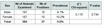 Table 1:Prevalence of mange mites in sheep by different sex factors
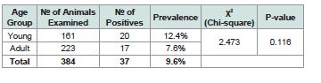 Table 2:Prevalence of mange mites in sheep by age group
 Table 3:Prevalence of mange mites in sheep among different body condition
scores
Table 3:Prevalence of mange mites in sheep among different body condition
scores
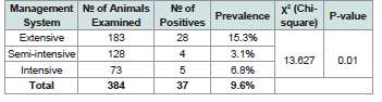 Table 4:Prevalence of mange mites in sheep by different management factors
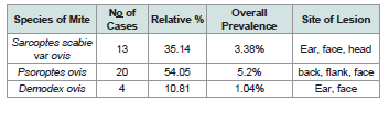 Table 5:Species of mange mites and distribution of lesions in the study sheep
Research Article
Prevalence of Mange Mites Infestation on Ovine in and Around Jimma Town, Southwest Ethiopia
Mekonen Baylie*
Fogera Woreda Animal Resources and Development Office, Woreta, South Gondar Zone, Amhara Regional State, Ethiopia
*Address for correspondence:Mekonen Baylie, Fogera Woreda Animal Resources and Development
Office, Woreta, South Gondar Zone, Amhara Regional State, Ethiopia E-mail: mekonenbaylie@gmail.com
Submission:05 February, 2024
Accepted:29 April, 2024
Published:04 May, 2024
Copyright: © 2024 Mekonen Baylie. This is an open access article
distributed under the Creative Commons Attribution License, which
permits unrestricted use, distribution, and reproduction in any medium,
provided the original work is properly cited.
Keywords:Mites; Mange; Prevalence; Sheep; Infestation; Skin Lesions;
Jimma
Abstract
A cross sectional study was conducted on 384 randomly selected
sheep in and around Jimma town, Oromia Regional State, Southwest
Ethiopia to assess the prevalence of ovine mange mites by laboratory
examination using skin scrappy. The results of the microscopical
examination of the skin scraping revealed that 37 of the sheep were
infested with mites with an overall prevalence of 9.6%. In this study,
three genus of mange mites were recorded that parasitized sheep;
namely, Sarcoptes, Psoroptes and Demodex with prevalence of 3.38%,
5.2% and 1.04% respectively, which were found on the back, shoulder,
tail, ear, face, ventral abdomen regions of the animals’ body. The
prevalence of the infestation was highest in sheep younger than two
years (12.4%) and the lowest in sheep with age older than two years
(7.6%). The prevalence of mange mites in male sheep was 9.1% and
10.2% in females. There were no statistically significant difference
(p>0.05) in the prevalence of mange mite infestation between the
different age and sex groups. The difference in the prevalence of
mange mites infestation in body condition score and management
practices was statistically significant (χ2=24.613, p=0.000), (χ2=13.627,
p=0.01, respectively). The dominant lesions of mange mites were
formations of nodules and crusts and also loss of hair and ragged
wool. Therefore, there should be immediate attention and control
interventions against the disease to cut the losses that hamper sheep
production and productivity in the study area.
Introduction
Sheep play a vital role as sources of meat, milk and wool for
smallholder keepers in different farming systems and agro-ecological
zones of Ethiopia [1]. Ethiopia is home to 23.6 million sheep [2] but
the immense potential numbers represent has yet to be realized due
to a multitude of factors. Ectoparasites are very common and widely
distributed in all agro-ecological zones in Ethiopia [3]. Ectoparasites
cause a wide range of health problems that confront the productivity
of sheep. Lice, sheep keds, ticks, fleas and mange mites are reported
to cause great pre-slaughter defects responsible for downgrading and
rejection of sheep skins. It is reported that 35% of sheep skin rejections
in Ethiopia are attributed to ectoparasites [4]. All these established
facts imply that ectoparasites pose serious economic losses to the
farmer, the tanning industry and the country as a whole [5].
In Oromia, there are an estimated 9,401,844 sheep, representing
36.2% of the national sheep population [2]. The Oromia region
supplies an estimated 32.9% of sheep skins to the central market
of the country. The export of processed and semi-processed skins
constitutes the second largest industry, next to coffee, in Ethiopia.
However, several recent reports indicate that over the last 10 years,
the quality of raw materials has deteriorated with an increase of skin
infestations associated with lice, sheep keds, ticks and mange mite [5].
Skin diseases caused by ectoparasites are among the major diseases
of sheep causing serious economic losses to small holder farmers, the
tanning industries and the countries as a whole. Such skin diseases
cause mortality, decrease production and reproduction and down
grading and rejection of skins. According to tanneries report, skin
diseases due to external parasites causes’ 35% sheep skin [6]. Mange
is a highly contagious skin disease caused by one or a combination
of several species of mites. Four genera of parasitic mites can cause
mange in sheep, namely Chorioptes species, Demodex species,
Psoroptes species and Sarcoptes species [7]. Hide and skin accounts
for 12-16% of the total volume of export from Ethiopia and though
hide and skins are important source of income, its contribution to the
national economy may far below [6]. There was no report of mange
mites’ infestation on sheep in and around Jimma town, Ethiopia.
Therefore, the objectives of the study were to estimate the prevalence
of mange mites of sheep in and around Jimma town, and to identify
the species of mange mites affecting sheep.
Materials and Methods
Study Area:
The study was conducted in and around Jimma town,
southwestern part of Ethiopia. Jimma town, the capital of Jimma
zone, is located in oromia regional administration 346 km southwest
of Addis Ababa at latitude of 7°40′N ′E and longitude of about 36°50′E
at elevation of 1,780 m above sea levels. The annual mean rainfall of
the area is about 1530 mm, and the minimum and maximum annual
mean temperature is 14.40C and 26.70C, respectively.
Study Design:
A cross sectional study design was carried out to estimate the
prevalence of mange mites infesting sheep. The prevalence of mange
mites, association of host related risk factors with the presence of mite
infestation were investigated. The host risk factors considered was
age, body condition, sex and management system. The examinations
of each animal were conducted by visual inspection and palpation of
skin lesions and by the eventual identification of ectoparasites. When
skin lesions were evidenced skins scrapping from suspected cases
of mange were collected. Mite identification was made according to
Taylor and Wall [8] and Wall and Sharer [9].
Study population:
The study animals were selected from the population of sheep in and around Jimma town.
Both sexes and all age groups were included in the study. Most samples were taken
from sheep that were brought to Jimma veterinary clinic for various
reasons. Records were also taken with regards to age, body condition,
sex and management.
Sample size and sampling method:
A simple random sampling method was used to select study animals. The sample size needed for
the study was calculated by using the formula given by Thrustfield
[10]. The study was considered 95% confidence interval and 5% level of precision.
n = 1.962 x Pexp (1-Pexp)/ d2
Where, n = required sample size
d = desired absolute precision at 95% confidence interval
Pexp = expected prevalence
The study considered expected prevalence of 50% for the sample size calculation, and hence, the sample size was 384. Therefore 384 sheep were examined in the study area.
Where, n = required sample size
d = desired absolute precision at 95% confidence interval
Pexp = expected prevalence
The study considered expected prevalence of 50% for the sample size calculation, and hence, the sample size was 384. Therefore 384 sheep were examined in the study area.
Study Methodology:
During clinical examination, age, sex, management and body
conditions of each sampled animal were recorded. Body condition
score of the animal was made as poor, medium and good; by
modifying the system described for sheep [11]. Poor body condition
score was given to sheep which was extremely thin, medium to those
with smooth and less prominent spinous process, transverse process
in which finger can be pushed and moderate depth loin muscle. Good
body condition score was given for the spinous process only stick up
very slightly; smooth, rounded and well covered transverse processes
and those having full loin muscle and very fat. Age categorization into
young (lamb) and adult was performed as described by [11] for sheep.
Accordingly, those sheep under 2 year were categorized as young and
the rest as adults.
Observation was undertaken on selected sheep to identify for
any lesions. From the area selected, skin scraping was taken using
universal bottle; and scraping area included the edge of a visible
lesion and the surrounding. After labeling the sample was transported
to Jimma University Veterinary Laboratory. Then the sample was
examined for mange mites under stereomicroscope. If during this
initial examination no mites were detected, further the samples were
heated on slide with a drop of 10% KOH. After allowing for 5-10
minutes with preparation to clear the debris, it was re-examined. The
species of mange mites were identified according to Taylor and Wall
[8] and Wall and Sharer [9].
Data Analysis:
The data collected were entered into Microsoft Excel spread sheets
and analyzed using SPSS (16.0) statistical software. The association
of mange mite infestation between body condition, management,
sex, and age of sheep were compared using Pearson Chi square test
(χ2) and a P-value of <0.05 was used as the determinant for level of
statistical significance.
Results
A prevalence study on mange mites of sheep was conducted
to determine the presence of the different mange mites and their
association with different host factors including age, sex, management
system and body condition scores. The current study revealed an
overall prevalence of 9.6% (n=37) from the total of 384 animals
examined.
Prevalence of mange mites infestation in sheep based on sex:
With regard to sex based prevalence of mange mites in this study,
both female and male sheep were infested with mange mites with an
overall prevalence of 9.6%, but the infestation in female was higher
than in male [Table 1]. However, the difference was not statistically
significant (p>0.05) [Table 1].
Prevalence of mange mites infestation in sheep based on age groups:
The prevalence study of mange mite in the different age groups
revealed a higher prevalence in young sheep compared with the
adult ones [Table 2]. Nonetheless, the difference was not statistically
significant (p>0.05) between the two age groups [Table 2].
Prevalence of ovine mange mites with regard to body condition scores:
With reference to prevalence of mange mites based on body
condition scores, it is found that poor body conditioned animals
were found to harbor more mange mite with a prevalence of 20.0%;
whereas medium body conditioned were 5.1% and good body
conditioned were 2.8% in prevalence [Table 3]. The difference in
the prevalence between poor, medium and good body conditioned
animals was statistically significant (p<0.05) [Table 3].
Prevalence of ovine mange mites on the basis of management systems:
The prevalence study of mange mite based on management
revealed that sheep under extensive animal management practice
were found to harbor more mange mite with a prevalence of 15.3%
where as sheep under semi-intensive had a 3.1% and those sheep
under intensive management had 6.8% prevalence [Table 4]. The
difference in the prevalence among the different management
practices was statistical significant (p<0.05) [Table 4].
Distribution of lesions and species of mange mites identified in sheep:
The predominant sites where mange lesions observed were the
face, back, head and ear area, and in general loss of hair and nodule
(solid dermal) formation were the dominant lesions recorded in the
present study [Table 5]. This study showed that sheep were infested
with three genera of mange mites namely. Sarcoptes scabiei was found
in 13 cases from a total of 37 infested cases with a relative percentage
of 35.14%. Psoroptes ovis was 20 cases with a relative percentage
54.05% and Demodex ovis was 4 cases with a relative percentage of
10.81% [Table 5].
Discussion
The present study revealed an overall prevalence of 9.6%. This
finding was higher than the previous findings in sheep documented
elsewhere in Ethiopia; 0.95% in Tigray region [12], 1.56% in and
around Mekele [13], and 2.1% in Sidama Zone [14]. However, the
finding of the current study was lower than the prevalence reported
in the Southern range land of Oromia, 14.64% in sheep [15]. This
discrepancy might be due to the different management status and the
use of acaricides and related control practices. This study revealed three
genera of mange mites namely, sarcoptes, psoroptes and demodex,
in the study area. The overall prevalence of sarcoptes was 3.38%.
However, sheep were reported to be rarely infested with sarcoptes
[16]. The lesion of Sarcoptes scabiei var ovis in sheep was observed
mostly around the ear, face and head areas and nodule formation was
the characteristic lesion recorded. Kassa [17] observed that, sarcoptic
mange if they occur in sheep in general they are frequently observed
in sparse hair. Kahn [16] also reported that sarcoptic mange mite
in sheep is very rare and if any, it is only seen in non-wooly areas.
In this study, psoroptes is the highest in prevalence (5.2%). This is
deviant from other findings where sarcoptes was reported as the most
prevalent species in [18].
In this study, Demodex ovis was recorded at the prevalence
of 1.04%. Similarly, several authors have reported nearly similar
prevalence, such as Numery [19] reported 1.36% in Kombolcha, north
eastern Ethiopia and also Shiferaw et al. [20] reported a prevalence of
demodecosis as 0.57% in sheep. According to Radostitis et al. [21],
demodectic mange is rare in sheep. The most important lesion was
nodule and crust formation found around the head and face. Similar
lesions of mange mites in sheep were reported by Chanie and Sirak
[6] and Kettle [22].
In this study, psoroptes ovis was recorded at the prevalence of
5.2%. This result is in line with the findings reported by Sertse [23]
and Shiferaw et al. [20]. The genus psoroptes was the most dominant
mange mite in sheep in other findings as well. For instance, psoroptes
in sheep in Baghdad province was reported by Currier et al. [24]
at 14% prevalence rate with Psoroptes mite and Tasawar et al. [25]
has reported 15% sheep scab prevalence in Scotland. The causes for
these variations may be due to the differences of the environmental
conditions and geographical areas. In the current study, the most
important lesion was nodule and crust formation that was found
on back, flank region. According to Pangui [26], high temperature,
humidity and sunlight favor mange mite infestation. The higher
temperature, humidity and sunlight which prevail in lowland and
midland may have accounted for the differences in prevalence.
Mange mite infestation is independent of age and sex [27]. Based
on our observation in this study, it is possible to conclude that age and
sex are not a predisposing risk factor to sheep with regard to mange
mite infestation. Similar result was reported by Shiferaw et al. [20]
where sex and age of the host animals are not determinant factors for
the prevalence variation. This finding was in agreement with previous
observation made elsewhere in Ethiopia. Yacob [28] and Kassaye and
Kebede [4] also had reported that sex has no significant effect on the
prevalence of mange mites. This may be due to the fact that both male
and female are exposed to the infestation as they are left to graze in
the same environment and no selective management is practiced in
the locality based on the sexes [29].This study showed difference in
mange mite infestation among different age groups being higher in
young age group. Kebede [4] and Shiferaw et al. [20] reported higher
prevalence of mange mite in young animals than the old age group.
The higher prevalence of mange in young than adult in the current
study is in line with the previous observations [30] and most probably
reflects the under-developed immunity in young animals.
Management practice attributed to the variation in the distribution
and abundance of mange mite in sheep. The management system can
contribute with regard to variations in prevalence of mange and the
existence of higher population of sheep in small area can facilitate the
infestation because the main way of transmission is intimate contact
[8]. However, the current study revealed that the sheep that managed
intensively has low prevalence than that was managed extensively,
this might be due to regular use of acaricides, ivermectin and proper
nutrition supplement practices in intensive management system.
In agreement with the current finding, Radostitis et al. [21] have
described that sarcoptic mange mite often go hand in hand with poor
feeding and general mismanagement.
Body condition attributed to the variation in the distribution and
abundance of mange mite in sheep. This highly significance difference
might be due to the fact that poor husbandry practices make
favorable condition for mange mite infestation. There are reports
elsewhere in agreement with this finding where animals which have
poor body condition appear most susceptible to infestation [8]. The
increased susceptibility of poor body conditioned animals to mange
is responsible for such difference [21]. In a similar report by Demissie
et al. [31], a prevalence of 15.3% mange mite infestation in poor body
condition was documented in selected sites of Amhara region. This
might be due to nutritional status, where well-fed animals can better
withstand parasitic infestation than animals on an inadequate diet
which can influence the level of immunity. In addition, the itching
and stress caused by the infestation can result in loss of appetite and
distraction from feeding and foraging activities of the animal which
might cyclically result in poor body condition. The burrowing and
feeding activities of S. scabie cause intense itching, inflammation, hair
loss and formation of crusts of exudates, loss of condition and death
[32]. Therefore, the economic impact of mange must be certainly high
judging from poor condition of affected animals, loss of affected skin,
deaths due to the disease and cost of treatments.
Conclusions and Recommendations
The study demonstrates that mange mite is among the most
important health constraints of sheep in and around Jimma, This
study demonstrated the occurrence and high prevalence of different
species of mange mites in sheep of all age groups, both sexes,
management practice and body conditions. The study revealed that
psoropte is the predominant mite in sheep followed by sarcoptes and
demodex species, hence requires immediate attention and control
interventions. In comparison with others studies, the present study
showed relatively higher mange mite infestation rate than the rest
of the country due to backward level of management, poor level of
awareness of farmers and weak animal health extension services
are believed to have contributed for wide spread distribution and
occurrence of mange mites in the study area leading to important
economic losses.
Therefore, from the above conclusion, the following
recommendations are forwarded:
• Control strategies should be instituted immediately taking in to account the impact of the parasite on the economy.
• The government should also give attention for control options besides application of acaricides spraying.
• In addition, further studies should be conducted taking in to account agro-ecological zones and mange mites’ distribution. And,
• Appropriate extension programs should be launched to create public awareness about the economic importance, treatments and its impact on skin quality.
Acknowledgements
The author is grateful to thank Elias Kebede (DVM, MSc), Jimma
University staffs and University of Gondar, Faculty of Veterinary
Medicine staffs.
References
Citation
Mekonen Baylie. Prevalence of Mange Mites Infestation on Ovine in and Around Jimma Town, Southwest Ethiopia. J Veter Sci Med. 2024;12(1): 1