Journal of Pediatrics & Child Care
Download PDF
Case Report
*Address for Correspondence: Sunil K. Jain, MD, FAAP, Department of Pediatrics/Division of Neonatology, University of Texas Medical Branch, 301 University Blvd, 6.104 Waverley Smith Pavilion, Galveston TX 77555-0526, USA, Tel: 409-772-2815; Fax: 409-772-0747; E-mail: skjain@utmb.edu
Citation: Bhargava V, Dasgupta S, Mahajan V, Jain SK. Silent Feto-Maternal Bleed as a Cause of Severe Neonatal Anemia. J Pediatr Child Care. 2016;2(1): 03.
Copyright © 2016 Jain, et al. This is an open access article distributed under the Creative Commons Attribution License, which permits unrestricted use, distribution, and reproduction in any medium, provided the original work is properly cited.
Journal of Pediatrics & Child Care | ISSN: 2380-0534 | Volume: 2, Issue: 1
Submission: 01 March, 2016 | Accepted: 19 April, 2016 | Published: 24 April, 2016
The next best step after eliminating the above etiologies is to determine the cause of blood loss in the infant. A high reticulocyte count, absence of tachycardia and hypotension make acute blood loss less likely in this case. This was a singleton pregnancy, with timely clamping of the umbilical cord ruling out twin to twin transfusion or blood loss secondary to delayed cord clamping as potential causes. Remaining etiologies for blood loss include fetal, feto-maternal and placental causes. An infant can bleed into several potential spaces including the subgaleal space, intra-abdominal and pelvic areas. However, a normal physical exam made these unlikely. The placenta was examined for signs of abruption, and was noted to be unremarkable for any blood clots or missing cotyledons.
Silent Feto-Maternal Bleed as a Cause of Severe Neonatal Anemia
Vidit Bhargava1, Soham Dasgupta1, Vasudha Mahajan1 and Sunil K Jain2*
- 1Department of Pediatrics, University of Texas Medical Branch Hospital, 300 University Boulevard, Galveston, Texas 77555, USA
- 2Department of Pediatrics/Division of Neonatology, University of Texas Medical Branch Hospital, 301 University Blvd, 6.104 Waverley Smith Pavilion, Galveston TX 77555-0526, USA
*Address for Correspondence: Sunil K. Jain, MD, FAAP, Department of Pediatrics/Division of Neonatology, University of Texas Medical Branch, 301 University Blvd, 6.104 Waverley Smith Pavilion, Galveston TX 77555-0526, USA, Tel: 409-772-2815; Fax: 409-772-0747; E-mail: skjain@utmb.edu
Citation: Bhargava V, Dasgupta S, Mahajan V, Jain SK. Silent Feto-Maternal Bleed as a Cause of Severe Neonatal Anemia. J Pediatr Child Care. 2016;2(1): 03.
Copyright © 2016 Jain, et al. This is an open access article distributed under the Creative Commons Attribution License, which permits unrestricted use, distribution, and reproduction in any medium, provided the original work is properly cited.
Journal of Pediatrics & Child Care | ISSN: 2380-0534 | Volume: 2, Issue: 1
Submission: 01 March, 2016 | Accepted: 19 April, 2016 | Published: 24 April, 2016
Abstract
Feto-maternal bleed is a common presentation during pregnancy that may have adverse consequences on the perinatal clinical course of the infant and mother alike. Anemia is a common presentation of an infant secondary to feto-maternal bleed. Large volume feto-maternal bleeds may lead to severe neonatal anemia in the absence of obvious maternal symptoms. Hence, a high index of suspicion must be maintained in the absence of other obvious causes of neonatal anemia. We describe a case of an infant with severe anemia secondary to a silent feto-maternal bleed associated with a normal appearing placenta on examination and the absence of obvious maternal symptoms.Introduction
Feto-maternal hemorrhage is defined as the entrance of fetal blood into the maternal circulation during pregnancy. Antenatal fetomaternal bleed is a pathological condition of variable significance. While, small volume bleeds are frequent and clinically insignificant; large volume bleeds are rare and can have adverse outcomes. We describe a case of a newborn infant presenting with severe anemia due to silent feto-maternal bleed. The diagnosis was complicated by vague maternal symptoms and completely normal appearance of the placenta on examination.Case Report
A male infant was born to a 29-year-old G1P1 mother at 39 weeks gestation via emergent caesarean section secondary to prolonged late decelerations. Birth weight was 2.8 kg. Apgar’s were 5/7/9 at 1/5/10 minutes. Initial examination was significant for an extremely pale appearance, mild-moderate respiratory distress requiring 2L30 % nasal cannula and a grade 2/6 systolic murmur best heard over the lower left sternal border. Capillary refill was 2 seconds, pulse rate 140-150 bpm and blood pressure was 63/32 mmHg. Mother had negative serologies including a negative screen for group B streptococcus. Mother complained of reduced fetal movements 24-48 hours prior to delivery with a corresponding biophysical profile of 4/10.Since the infant appeared pale, complete blood count (CBC) was obtained which was significant for a white cell count of 54100/μl, hemoglobin (Hgb) of 4.1 g/dl (19.3 ± 2.2 g/dl), hematocrit (Hct) of 13.7% (61 ± 7.4 %), platelets of 206,000/μl and an immature to total neutrophil cell (I/T) ratio of 0.31. Reticulocyte count was elevated at 8.9% (3.2 ± 1.4). Maternal blood type was O positive and baby’s blood type was B negative with a negative direct Coombs test. There was no evidence of abruption on examination of the placenta. The placenta was free of any blood clots, the amniotic fluid was clear and no clots were noted in the uterus during cesarean section. Blood culture was obtained and the baby was started on prophylactic antibiotics secondary to the abnormal white cell count raising concerns for sepsis. Transcutaneous bilirubin was measured at 0.8 mg/dl and later a serum bilirubin showed an unconjugated bilirubin of 0.5 mg/dl and conjugated bilirubin of 0.0 mg/dl. On physical exam, baby had a normal anterior fontanelle without any signs of bulging and a soft non-distended, non-tender abdomen with normal bowel sounds. 20 ml/kg of packed red blood cells was emergently transfused. Blood was transfused over 4 hours in 2 aliquots without any complications. CBC post transfusion showed a Hgb of 10.9 g/dl and a Hct of 31.6%, The heart murmur was no longer audible post transfusion. The baby continued to be hemodynamically stable post transfusion. Hemoglobin electrophoresis was sent and it returned normal results. Further maternal history and an additional bedside test revealed the diagnosis.
Discussion
Anemia is defined as hematocrit or hemoglobin concentration < 2SD below age specific mean [1]. The etiology can be broadly classified into a) decreased production of RBC’s, b) increased destruction of RBC’s or c) increased blood loss in the fetus [2].Establishing hemodynamic stability is of paramount importance in a newborn presenting with extreme pallor. Once this is achieved, the next step should be confirming suspected anemia. In this case the initial Hgb/Hct after birth was found to be extremely low, consistent with severe anemia. The initial elevated white count was likely due to an erythroblastic reaction secondary to severe anemia and not due to sepsis. Cultures were negative and the white count normalized within 2 days of transfusion [3]. Anemia in a newborn can have multiple etiologies. A stepwise approach to diagnosis based on clinical acumen results in minimal investigations and early, effective interventions. Such an approach was followed to establish the diagnosis in this patient.
Reticulocyte count is a simple yet effective marker for differentiating between acute and chronic blood loss. A high reticulocyte count can be seen secondary to bone marrow compensation in the events of hemolysis (increased RBC destruction) or chronic blood loss. On the other hand, a low or normal reticulocyte count implies decreased RBC production secondary to bone marrow failure or due to an acute loss of blood. The high reticulocyte count in our patient made bone marrow suppression secondary to congenital or acquired causes highly unlikely.
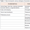
Hemolysis can be caused by several etiologies (Table 1). Hemolysis leads to unconjugated hyper-bilirubinemia in neonates. A transcutaneous bilirubin and later a serum bilirubin were noted to be low (0.8 mg/dl and 0.5 mg/dl respectively) making hemolysis less likely.
The next best step after eliminating the above etiologies is to determine the cause of blood loss in the infant. A high reticulocyte count, absence of tachycardia and hypotension make acute blood loss less likely in this case. This was a singleton pregnancy, with timely clamping of the umbilical cord ruling out twin to twin transfusion or blood loss secondary to delayed cord clamping as potential causes. Remaining etiologies for blood loss include fetal, feto-maternal and placental causes. An infant can bleed into several potential spaces including the subgaleal space, intra-abdominal and pelvic areas. However, a normal physical exam made these unlikely. The placenta was examined for signs of abruption, and was noted to be unremarkable for any blood clots or missing cotyledons.
Feto-maternal bleed is seen in about 96% of pregnancies [4], however this is often less than 1 ml. Significant feto-maternal bleed can lead to severe anemia as seen in our patient and often may lead to fetal death in utero. The diagnosis of a feto-maternal bleed can be made by a simple bedside test known as the Kleihauer-Betke (KB) test [5]. In this case, the KB test obtained within 1 hour of delivery, showed a fetal cell count of 90 in 2000, representing 150 ml fetal blood which explains the severe anemia seen in this patient. On retrospective questioning, the mother confirmed that she had cramping pain a few days prior to delivery which was different in character as compared to her regular contractions. This, in combination with the decreased fetal movements is suggestive of a silent feto-maternal bleed. A high index of suspicion is required in order to diagnose silent fetomaternal bleed since these patients are frequently asymptomatic with a soft and non-tender uterus; however the associated hemorrhage may be excessive and the fetal mortality is high [6].
Conclusion
In conclusion, neonatal anemia has an extensive differential diagnosis. However, clinical judgment and reasoning can help narrow down the causes and thus the laboratory tests that are needed to establish diagnosis.References
- Kett JC (2012) Anemia in infancy. Pediatr Rev 33.
- Widness JA (2008) Pathophysiology of anemia during the neonatal period, including anemia of prematurity. Neoreviews 9: e520.
- Dua V, Jain V, Yadav SP, Sachdeva A (2012) Interpretation of the complete blood count. In: Sachdeva A (Ed). Pediatric practical hematology, 2nd edition. Jaypee Brothers Medical Publishers Inc., Panama City, Panama, pp. 1-11.
- Sebring ES, Polseky HF (1990) Fetomaternal hemorrhage: incidence, risk factors, time of occurrence and clinical effects. Transfusion 30: 344-357.
- Kleihauer E, Braun H, Betke K (1957) Demonstration von fetalem hemoglobin in den erythrocyten eines blutausstrichs. Klin Wochenschr 35: 637-638.
- Notelovitz M (1974) Silent abruption of the posteriorly inserted placenta. S Afr Med J 48: 93-95.


