

Journal of Cardiobiology
Download PDF
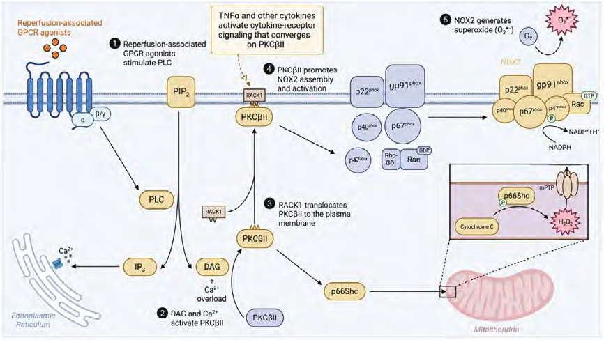 Figure 1:Proposed PKCβII-mediated ROS signaling during myocardial ischemia–reperfusion, showing convergent cytokine-receptor and GPCR signaling to PKCβII, NOX2 activation, and mitochondrial ROS production.
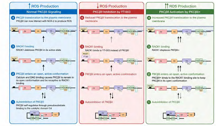 Figure 2:Proposed model comparing basal PKCβII signaling, YT-003–mediated inhibition of RACK1-dependent PKCβII localization, and the predicted effects of PKCβII+ on PKCβII activation and ROS production.
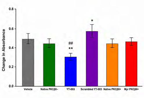 Figure 3:Superoxide production in PMA-stimulated isolated rat PMNs,
showing that YT-003 significantly reduced superoxide release compared with
vehicle and scrambled YT-003 controls; data are mean ± SEM, *p < 0.05 and
**p < 0.01 versus vehicle control, ##p < 0.01 versus scrambled YT-003, n =
6 independent experiments.
Figure 3:Superoxide production in PMA-stimulated isolated rat PMNs,
showing that YT-003 significantly reduced superoxide release compared with
vehicle and scrambled YT-003 controls; data are mean ± SEM, *p < 0.05 and
**p < 0.01 versus vehicle control, ##p < 0.01 versus scrambled YT-003, n =
6 independent experiments.
For the hypoxia–reoxygenation assay, HUVECs were placed in a modular incubator chamber and exposed to hypoxic conditions (1% O₂, 5% CO₂, and balanced N₂) for 24 h. Cells were then returned to normoxic conditions for 24 h, with drug treatment administered at the onset of reoxygenation. Cell viability was assessed at the end of the reoxygenation period using a WST-8 tetrazolium-based colorimetric assay, and absorbance was measured at 450 nm using a microplate reader. Results were normalized to normoxic control conditions.
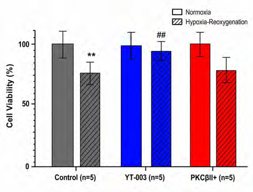 Figure 4:HUVEC viability under normoxic conditions or following hypoxia–
reoxygenation with YT-003 or a myristoylated PKCβII activator, showing
significant preservation of viability by YT-003 compared with the hypoxia–
reoxygenation control; **p = 0.004 versus normoxic control and ##p = 0.0087
versus hypoxia–reoxygenation control.
Figure 4:HUVEC viability under normoxic conditions or following hypoxia–
reoxygenation with YT-003 or a myristoylated PKCβII activator, showing
significant preservation of viability by YT-003 compared with the hypoxia–
reoxygenation control; **p = 0.004 versus normoxic control and ##p = 0.0087
versus hypoxia–reoxygenation control.
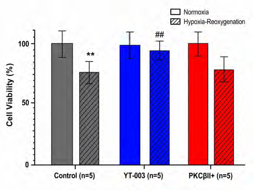 Figure 5:Myocardial infarct size in isolated rat hearts following ex vivo
ischemia–reperfusion; data are mean ± SEM, and YT-003 significantly
reduced infarct size versus control (*p = 0.026) and scrambled YT-003
(#p = 0.019).
Figure 5:Myocardial infarct size in isolated rat hearts following ex vivo
ischemia–reperfusion; data are mean ± SEM, and YT-003 significantly
reduced infarct size versus control (*p = 0.026) and scrambled YT-003
(#p = 0.019).
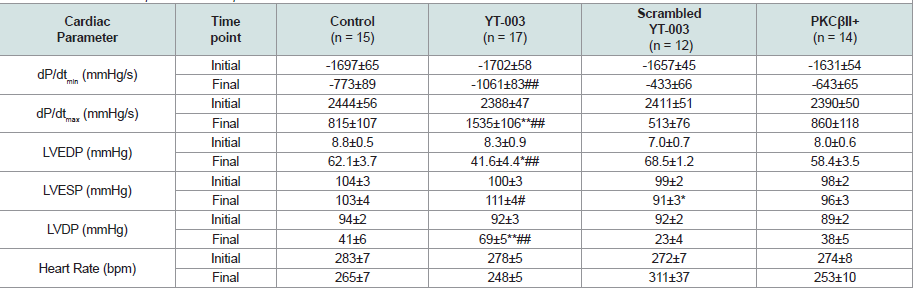 Table 1:Cardiac functional parameters at baseline and after 50 min of reperfusion in an ex vivo isolated rat-heart model of myocardial ischemia–reperfusion, showing that YT-003 significantly improved left ventricular function compared with control and scrambled YT-003; data are mean ± SEM, with *p < 0.05 and **p <0.01 versus control and #p < 0.05 and ##p < 0.01 versus scrambled YT-003.
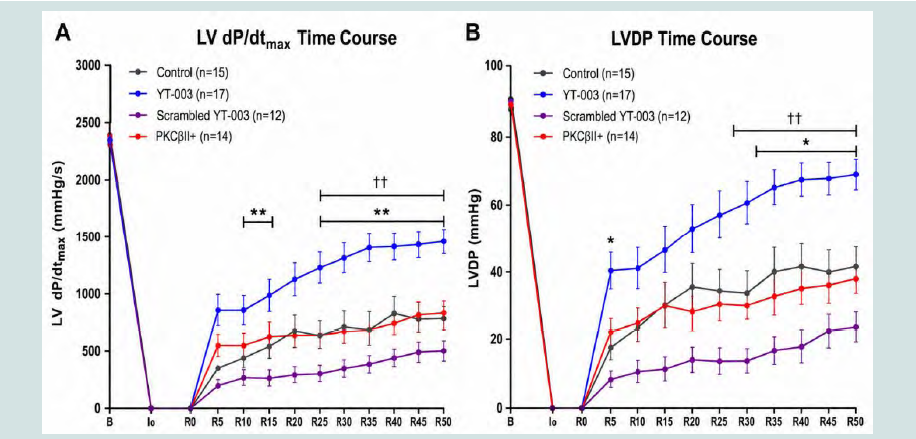 Figure 6:Left ventricular functional recovery in isolated rat hearts following ex vivo ischemia–reperfusion, showing that YT-003 improved dP/dtmax (A) and LVDP (B) during reperfusion compared with control and PKCβII+; B, baseline; I0, onset of ischemia; R0, onset of reperfusion; *p < 0.05 and **p <
0.01 versus control; ††p < 0.01 versus PKCβII+.
Figure 6:Left ventricular functional recovery in isolated rat hearts following ex vivo ischemia–reperfusion, showing that YT-003 improved dP/dtmax (A) and LVDP (B) during reperfusion compared with control and PKCβII+; B, baseline; I0, onset of ischemia; R0, onset of reperfusion; *p < 0.05 and **p <
0.01 versus control; ††p < 0.01 versus PKCβII+.
Building on these findings, our laboratory subsequently evaluated whether additional lipid conjugation could further improve peptide delivery and biological activity. Our previous studies demonstrated that dual conjugation of PKC-targeting peptides with myristic acid (Myr) and Tat significantly improved intracellular uptake and cardioprotective efficacy compared with single-conjugated peptides [58,61]. As a result, we developed a formulation of our PKCβII inhibitor, YT-004, that is conjugated to both myristic acid and Tat. In ex vivo rat hearts, YT-004 significantly reduced infarct size at femtomolar concentrations, while in vivo porcine myocardial ischemia–reperfusion models showed cardioprotection at doses as low as 20 ng/kg, corresponding to approximately 100 pM circulating drug levels [62,63]. These findings suggest that dual-conjugated PKCβII inhibitors may achieve potent biological effects at substantially lower concentrations than earlier-generation compounds.
Future studies will evaluate YT-004 across in vitro, ex vivo, and in vivo systems to further define its therapeutic efficacy and translational potential. Planned investigations include testing YT-004 in a clinically relevant porcine myocardial ischemia–reperfusion model involving regional ischemia followed by reperfusion and extended post-injury follow-up to assess chronic recovery. Longitudinal assessment will include left ventricular ejection fraction, hemodynamic performance, and circulating biomarkers of myocardial injury, with infarct size and post-injury remodeling quantified at study completion. Additional studies will characterize pharmacokinetics, tissue distribution, and dosing parameters to optimize therapeutic exposure and determine the durability of treatment effects.
Research Article
Targeting Protein Kinase C Beta II to Reduce Myocardial Ischemia–Reperfusion Injury
Nair A1, Beale MA1, Dean TC1, Singh SG1, Michaels M1, Baker A1, Madison IT1, Lloyd M1, Castro J1, Dang J1, Metellus D1, Melnik J1, Le A1, Chen Q1, Barsotti R1, and Young LH1,2*
1Department of Bio-Medical Sciences, Philadelphia College of Osteopathic
Medicine (PCOM), Philadelphia, PA 19131, USA
2Young Therapeutics, LLC, Philadelphia, PA 19152, USA
2Young Therapeutics, LLC, Philadelphia, PA 19152, USA
*Address for Correspondence:Lindon H. Young, Department of Bio-Medical Sciences Philadelphia College of Osteopathic Medicine 4170 City Avenue Philadelphia, PA 19131 USA. E-mail Id: lindonyo@pcom.edu
Submission: 29 May, 2026
Accepted: 13 August, 2026
Published: 18 August, 2026
Copyright: © 2026 Nair A, et al. This is an open access article distributed
under the Creative Commons Attribution License, which permits
unrestricted use, distribution, and reproduction in any medium, provided the
original work is properly cited.
Keywords:Myocardial Infarction; Cardioprotection; Yt-003; Reactive Oxygen
Species; Nicotinamide Adenine Dinucleotide Phosphate Oxidase 2; Receptor for
Activated C Kinase 1 (Rack1).
Abstract
Myocardial infarction (MI) remains a leading cause of morbidity and mortality
worldwide and is most commonly caused by coronary artery occlusion. Although
restoration of coronary blood flow is essential to preserve viable myocardium,
reperfusion paradoxically induces additional injury through mechanisms involving
reactive oxygen species (ROS) generation and inflammation, collectively termed
myocardial ischemia–reperfusion (MIR) injury. Protein kinase C beta II (PKCβII) has
emerged as a potential therapeutic target in MIR because of its role in regulating
oxidative and inflammatory signaling pathways; however, the relative effects of
PKCβII activation versus inhibition remain incompletely defined. Accordingly, we
evaluated a selective cell-permeable PKCβII inhibitor (YT-003) and a selective PKCβII
activator using complementary in vitro and ex vivo MIR models.
Rat polymorphonuclear leukocytes (PMNs) were pretreated with selective PKCβII modulators and chemically stimulated to generate superoxide (SO) to assess ROSgenerating capacity. Human umbilical vein endothelial cells (HUVECs) underwent hypoxia and reoxygenation, with treatment given at the onset of reoxygenation, and viability was assessed. Isolated rat hearts underwent global ischemia followed by reperfusion in a Langendorff preparation, with treatment administered at reperfusion onset; cardiac functional recovery and infarct size were measured.
YT-003 significantly reduced PMN-derived SO release, improved HUVEC survival after hypoxia–reoxygenation, decreased myocardial infarct size, and increased left ventricular functional recovery compared with controls. In contrast, the selective PKCβII activator did not significantly alter outcomes in any of the experimental models.
These findings indicate that selective PKCβII inhibition with YT-003 is protective in complementary in vitro and ex vivo MIR models. The activator data indicate a lack of efficacy for this particular peptide, rather than excluding a protective role for PKCβII activation more generally. These results are hypothesis-generating and require confirmation in clinically relevant in vivo models before the therapeutic potential of PKCβII inhibition after MI can be defined.
Rat polymorphonuclear leukocytes (PMNs) were pretreated with selective PKCβII modulators and chemically stimulated to generate superoxide (SO) to assess ROSgenerating capacity. Human umbilical vein endothelial cells (HUVECs) underwent hypoxia and reoxygenation, with treatment given at the onset of reoxygenation, and viability was assessed. Isolated rat hearts underwent global ischemia followed by reperfusion in a Langendorff preparation, with treatment administered at reperfusion onset; cardiac functional recovery and infarct size were measured.
YT-003 significantly reduced PMN-derived SO release, improved HUVEC survival after hypoxia–reoxygenation, decreased myocardial infarct size, and increased left ventricular functional recovery compared with controls. In contrast, the selective PKCβII activator did not significantly alter outcomes in any of the experimental models.
These findings indicate that selective PKCβII inhibition with YT-003 is protective in complementary in vitro and ex vivo MIR models. The activator data indicate a lack of efficacy for this particular peptide, rather than excluding a protective role for PKCβII activation more generally. These results are hypothesis-generating and require confirmation in clinically relevant in vivo models before the therapeutic potential of PKCβII inhibition after MI can be defined.
Abbreviations
Diacylglycerol (DAG); Dimethyl sulfoxide (DMSO); Endothelial
nitric oxide synthase (eNOS); Human umbilical vein endothelial
cells (HUVECs); Ischemia–reperfusion (I/R); Left ventricular
developed pressure (LVDP); Left ventricular end-diastolic pressure
(LVEDP); Left ventricular end-systolic pressure (LVESP); Mitochondrial
permeability transition pore (mPTP); Myocardial infarction
(MI); Myocardial ischemia–reperfusion (MIR); Myristic acid
(Myr); Myristic acid- and Tat-conjugated PKCβII inhibitor (YT-
004); Myristoylated PKCβII activator, N-Myr-SVEIWD (PKCβII+);
Myristoylated PKCβII inhibitor, N-Myr-SLNPEWNET (YT-003);
Nicotinamide Adenine Dinucleotide Phosphate Oxidase 2 (NOX-
2); Nitric oxide (NO); Phorbol 12-myristate 13-acetate (PMA);
Polymorphonuclear leukocytes (PMNs); Protein kinase C beta II
(PKCβII); Rate of decrease in left ventricular pressure (dP/dtmin);
Rate of increase in left ventricular pressure (dP/dtmax); Reactive
oxygen species (ROS); Receptor for activated C kinase 1 (RACK1);
Standard error of the mean (SEM); Superoxide (SO); Trans-activator
of transcription peptide (Tat); Tumor necrosis factor-alpha (TNFα)
Introduction
Myocardial infarction (MI) remains a leading cause of morbidity
and mortality worldwide and represents a substantial healthcare
burden [1-3]. In the United States alone, more than 750,000 MIs occur
annually, often resulting in significant, long-term cardiovascular
complications among survivors [4,5]. MI is most commonly caused
by partial or complete occlusion of a coronary artery. Current clinical
interventions focus on rapidly restoring coronary blood flow to
salvage ischemic myocardium. Although reperfusion limits overall
infarct size, it paradoxically induces additional tissue injury known
as myocardial ischemia–reperfusion (MIR) injury. MIR injury can
account for up to 50% of total final infarct size, highlighting it as a
critical and under-addressed therapeutic target [6,7]. Despite its
clinical significance, only one FDA-approved therapy currently exists
for MIR injury [8].
Protein kinase C beta II (PKCβII), a calcium and diacylglycerol
(DAG)-dependent serine/threonine kinase, emerged as a potential
mediator of MIR injury through its established roles in inflammatory
signaling and cellular stress responses [6,7,9-13]. PKCβII expression
is elevated in patients with ischemic cardiomyopathy, and
pharmacologic inhibition of PKCβII has been shown to improve
cardiac function in multiple experimental models of MIR injury
[14-18]. Prior work has suggested that low-level, long-term PKCβ
expression during adulthood may improve postischemic recovery
in mice [19]. However, whether acute selective PKCβII activation
during reperfusion is protective, neutral, or harmful remains
incompletely defined. The present study addresses this question by
directly comparing the effects of PKCβII activation and inhibition
within identical MIR injury models.
Our laboratory has developed a selective PKCβII inhibitor, YT-003
(Young Therapeutics), which blocks PKCβII-mediated production
of reactive oxygen species (ROS). ROS play a central role in MIR
injury by promoting oxidative damage, impairing vascular function,
and amplifying inflammatory signaling pathways [20,21]. PKCβII is
a known upstream regulator of ROS generation during ischemia–
reperfusion. PKCβII activation is initiated early during reperfusion by
the release of pro-inflammatory cytokines, including tumor necrosis
factor-α (TNFα) [22]. TNFα and other pro-inflammatory cytokines
bind to their respective cytokine receptors and activate intracellular
signaling pathways that converge on PKCβII activation. In parallel,
reperfusion-associated agonists acting through G-protein–coupled
receptors stimulate phospholipase C, increasing intracellular Ca²⁺
and diacylglycerol (DAG), which further promote PKCβII activation
[Figure 1] [23-26]. Activated PKCβII binds to the receptor for
activated C kinase 1 (RACK1) and is translocated to the plasma
membrane, where it phosphorylates p47phox, promoting assembly of
NADPH oxidase 2 (NOX-2) [27-29]. NOX-2 subsequently generates
superoxide radicals that contribute directly to MIR injury.
In addition to regulating NOX-2–dependent ROS generation,
PKCβII also contributes to mitochondrial oxidative stress
during reperfusion [Figure 1]. Following activation, PKCβII can
phosphorylate the redox adaptor protein p66Shc, causing it to
enter the mitochondria where it facilitates the oxidation of reduced
cytochrome c and generation of hydrogen peroxide [30-32]. This
increase in mitochondrial ROS has been linked to opening of
the mitochondrial permeability transition pore (mPTP), loss of
mitochondrial integrity, and initiation of cell injury pathways
associated with ischemia–reperfusion damage [33-35]. Together,
these findings suggest that PKCβII amplifies reperfusion injury
through both NOX-2 signaling and mitochondrial ROS production.
[Figure 2] summarizes basal PKCβII signaling and the divergent
effects of YT-003 and our PKCβII activator. YT-003 is designed
to bind RACK1 and impair RACK1-dependent localization of
activated PKCβII to membrane-associated signaling complexes,
thereby limiting downstream ROS production [Figure 2]. In prior
work, YT-003 significantly and concentration-dependently reduced
superoxide release from isolated rat polymorphonuclear leukocytes
(PMNs), increased nitric oxide production in rat aortic segments, and
improved functional recovery in an ex vivo rat heart MIR model [12].
Our PKCβII activator was designed as a pseudo-RACK sequence to
promote an active-like PKCβII conformation that facilitates RACK1
interaction and membrane-associated localization [Figure 2] [29].
Based on this mechanistic framework and prior findings, we
predicted that YT-003 would replicate our previously observed
cardioprotective effects by significantly decreasing PMN SO release,
improving endothelial cell survival after hypoxic stress, reducing
cardiac infarct size, and improving left ventricular function in an
ex vivo rat heart MIR model [12]. Conversely, we hypothesized that
PKCβII activation would produce the opposite effects, resulting
in increased PMN superoxide release, decreased endothelial cell
survival, larger infarct size, and impaired functional recovery.
Methods
All experimental protocols were performed in accordance with
the institutional policies of the Philadelphia College of Osteopathic
Medicine, and all animal procedures were additionally approved by
the Institutional Animal Care and Use Committee.
Drug Conjugation:
Our PKCβII inhibitor peptide (SLNPEWNET) and PKCβII
activator peptide (SVEIWD) were used to assess the effects of
PKCβII inhibition and activation, respectively. Each peptide was
tested in its native, unconjugated form and conjugated to myristic
acid (Myr), an endogenous fatty acid that enhances cell membrane
permeability, to determine whether myristoylation improved
intracellular delivery. YT-003 denotes the myristoylated PKCβII
inhibitor N-Myr-SLNPEWNET. A scrambled version of YT-003
(N-Myr-WNPESLNTE) was synthesized and used as a control. In
the figures and tables, the myristoylated PKCβII activator (N-Myr-
SVEIWD) is designated PKCβII+, the unconjugated activator
peptide as Native PKCβII+, and the unconjugated inhibitor peptide
as Native PKCβII−; the myristoylated inhibitor is designated YT-003
throughout. All peptides were tested at a final concentration of 20
μM in 0.4% dimethyl sulfoxide (DMSO). The PMN assay evaluated
the native and myristoylated PKCβII inhibitor and activator peptides,
as well as scrambled YT-003. The HUVEC assay evaluated YT-003
and the myristoylated PKCβII activator (PKCβII+), whereas the
isolated-heart experiments evaluated YT-003, scrambled YT-003,
and PKCβII+. Previous concentration–response studies of YT-003
in PMN superoxide (SO) release and ex vivo rat MIR hearts were
reported by Omiyi et al. (2005) and Lipscombe et al. (2015) [12,16].
Rat PMN SO Release:
Male Sprague-Dawley rats (350–400 g, Charles River Laboratories,
Springfield, MA) were housed in a 12-h light/12-h dark cycle in a
temperature-controlled room. Animals had ad libitum access to food
and water. All procedures were conducted in accordance with animal
handling protocol A21-002. Rats were placed under anesthesia with
2.5% isoflurane and injected intraperitoneally with 16 mL of 0.5%
glycogen to induce rat peritonitis and subsequent PMN recruitment.
After 18 h, rats were re-anesthetized with 2.5% isoflurane, and PMNs
were harvested by peritoneal lavage as previously described [36].
PMN yield and viability were assessed by trypan blue exclusion.
PMA-induced superoxide release was measured spectrophotometrically using a previously published ferricytochrome c reduction assay [37]. For each assay, a 900-μL reaction mixture was
prepared containing 450 μL of 0.25% ferricytochrome c solution, 5
× 10⁶ PMNs, 2 μL of the designated treatment, and molecular-grade
water. Dimethyl sulfoxide (DMSO, 0.4%) was used as the vehicle
control. Reaction mixtures were transferred to cuvettes, placed in
a spectrophotometer, and incubated for 15 min at 37 °C. Baseline
absorbance was recorded at 550 nm. PMNs were then activated by
adding 100 μL of a 100 nM phorbol 12-myristate 13-acetate (PMA)
stock to a final concentration of 10 nM, and absorbance was recorded
every 30 s for 390 s. Three treatment groups and one vehicle control
were analyzed simultaneously. Only assays in which vehicle-control
absorbance increased by greater than 0.300 absorbance units were
included in the final analysis.
HUVEC Hypoxia–Reoxygenation Assay:
Human umbilical vein endothelial cells (HUVECs) from a single
donor were obtained from Lonza (Walkersville, MD) and cultured in
endothelial cell growth medium on 0.2% gelatin-coated 96-well plates.
Culture medium was supplemented with 2% fetal bovine serum and
EBM-2 BulletKit components (Lonza, CC-3162). Cells were passaged
no more than 5 times and grown to approximately 80% confluence
under normoxic conditions (21% O₂, 5% CO₂, 85% relative humidity,
37 °C). Culture medium was replaced every 48 h.For the hypoxia–reoxygenation assay, HUVECs were placed in a modular incubator chamber and exposed to hypoxic conditions (1% O₂, 5% CO₂, and balanced N₂) for 24 h. Cells were then returned to normoxic conditions for 24 h, with drug treatment administered at the onset of reoxygenation. Cell viability was assessed at the end of the reoxygenation period using a WST-8 tetrazolium-based colorimetric assay, and absorbance was measured at 450 nm using a microplate reader. Results were normalized to normoxic control conditions.
Rat MIR Model:
Male Sprague-Dawley rats (275–325 g, Charles River Laboratories,
Springfield, MA) were housed in identical conditions to those
described for the PMN SO experiments. Rats were anesthetized via
an intraperitoneal injection of pentobarbital (60 mg/kg) and heparin
(1000 units). The heart was subsequently excised and immediately
perfused with Krebs buffer at 37 °C at a constant pressure of 80
mmHg using a Langendorff perfusion apparatus. Baseline cardiac
parameters were recorded during a 15-min stabilization period by
placing a SPR-425 catheter (Millar Instruments, Inc., Houston, TX)
into the left ventricle. The heart was then subjected to 30 min of global
ischemia by stopping perfusion, followed by 50 min of reperfusion
during which perfusion was restored. The designated drug treatment
or vehicle control (0.4% DMSO) was delivered during the first 5 min
of reperfusion at a rate of 1 mL/min. Heart rate, left ventricular endsystolic
pressure (LVESP), left ventricular end-diastolic pressure
(LVEDP), the rate of increase in left ventricular pressure (dP/dtmax),
and the rate of decrease in left ventricular pressure (dP/dtmin) were
recorded at 5-min intervals throughout the reperfusion period. Data
were acquired and stored using a PowerLab/8Sp data acquisition
system (AD Instruments, Colorado Springs, CO). Left ventricular
developed pressure (LVDP) was calculated as the difference between
LVESP and LVEDP.
At the conclusion of the reperfusion period, the heart was
removed from the Langendorff apparatus and placed in a −20 °C
freezer for 30 min. The hearts were then sectioned perpendicular
to their long axis into seven 2-mm slices and incubated for 5 min
at 37 °C in 1% 2,3,5-triphenyltetrazolium chloride dissolved in 0.2
M Tris buffer (pH 7.4). Following incubation, the slices were fixed
in 4% paraformaldehyde. Viable tissue stained red, whereas infarcted
tissue appeared pale. Percent infarction was determined by separating
the infarcted from the viable tissue and calculating the ratio of the
infarcted tissue weight to the total slice weight.
Statistical Analysis:
All data are presented as mean ± SEM. Normally distributed
data were analyzed using ANOVA followed by post hoc pairwise
comparisons using the Games-Howell test. Non-normally
distributed data were analyzed using the Kruskal-Wallis test followed
by Wilcoxon rank-sum pairwise comparisons with Bonferroni
correction. Values of p < 0.05 were considered statistically significant.
Statistical symbols, significance thresholds, and the corresponding
comparison groups are defined in the individual figure legends and
table footnote.
Results
PKCβII Inhibition Reduces Superoxide Release in Isolated PMNs
Superoxide (SO) production was measured in isolated rat
polymorphonuclear leukocytes (PMNs) to determine whether
PKCβII activation or inhibition alters reactive oxygen species
generation after PMA stimulation [Figure 3]. Superoxide release is
expressed as the change in absorbance at 550 nm recorded 390 s after
PMA stimulation. Neither the native PKCβII inhibitor (0.43 ± 0.02, n
= 22) nor the native PKCβII activator (0.43 ± 0.04, n = 15) significantly
altered SO release compared to the vehicle control (0.46 ± 0.02, n
= 74). In contrast, PMNs treated with YT-003 exhibited a marked
reduction in SO release at 390 s (0.30 ± 0.02, n = 27). This reduction
was significant compared with vehicle control (p = 0.000012), the
native PKCβII inhibitor (p = 0.0043), and scrambled YT-003 (p =
0.00028). The myristoylated PKCβII activator did not significantly
alter SO production (0.44 ± 0.02, n = 26). Notably, treatment with
scrambled YT-003 resulted in a significant increase in SO release
(0.58 ± 0.05, n = 22) relative to vehicle-treated controls (p = 0.016).
KCβII Inhibition Improves Endothelial Cell Viability in a Hypoxia–Reoxygenation Model:
Endothelial cell survival following ischemic stress was assessed
using a hypoxia–reoxygenation model in human umbilical vein
endothelial cells (HUVECs). For each condition, n = 5 represents five
measurements obtained in five independent experiments. Exposure
to 24 h of hypoxia followed by 24 h of reoxygenation significantly
reduced HUVEC viability to 79.0 ± 8.7% of the normoxic control (n
= 5; p = 0.004; (Figure 4). Administration of YT-003 at the onset of
reoxygenation significantly improved endothelial viability (94.7 ±
6.8%, n = 5, p = 0.0087). In contrast, treatment with the myristoylated
PKCβII activator did not significantly improve cell viability, which
remained approximately 81.4 ± 9.7% (n = 5, p = 0.81).
PKCβII Inhibition Reduces Infarct Size in an Ex Vivo Rat MIR Model:
To determine whether PKCβII modulation influences myocardial
tissue injury, isolated rat hearts were subjected to 30 min of global
ischemia followed by 50 min of reperfusion in an ex vivo myocardial
ischemia–reperfusion model. Hearts treated with YT-003 at the onset
of reperfusion exhibited a significantly smaller infarct size (13.0 ±
1.8%, n = 17) compared to DMSO-treated controls (24.3 ± 3.5%, n
= 15, p = 0.026) and hearts treated with scrambled YT-003 (22.0 ±
1.9%, n = 12, p = 0.019) (Figure 5). In contrast, treatment with the
myristoylated PKCβII activator did not significantly alter infarct size
(21.4 ± 2.6%, n = 14, p = 1.0).
PKCβII Inhibition Improves Left Ventricular Functional Recovery During Reperfusion:
YT-003 treatment significantly improved left ventricular
functional recovery during reperfusion. YT-003 significantly
increased dP/dtmax compared with vehicle control from 10 min to
the end of reperfusion, except at 20 min (Figure 6A). YT-003 also
significantly increased dP/dtmax compared to scrambled YT-003
from 10 min to the end of reperfusion and compared to the PKCβII
activator from 25 min to the end of reperfusion. In addition, YT-003
significantly increased LVDP compared to vehicle control at 5 min
and from 30 min to the end of reperfusion, to scrambled YT-003 from
5 min to the end of reperfusion, and to the PKCβII activator from 25
min to the end of reperfusion (Figure 6B). Interestingly, scrambled
YT-003 significantly reduced LVDP compared with control between
20 and 40 min of reperfusion. The myristoylated PKCβII activator
did not significantly improve dP/dtmax, LVDP, or any other cardiac
parameters compared to vehicle control under the conditions
examined [Figure 6] [Table 1]. Heart rate did not differ significantly
between any groups [Table 1].
Discussion
Summary of Major Findings:
Our study compared the effects of PKCβII activation and
inhibition on MIR injury. YT-003 significantly decreased SO
release from isolated rat PMNs and improved HUVEC viability in
a hypoxia–reoxygenation assay. In an ex vivo rat heart MIR model,
YT-003 significantly reduced infarct size and significantly improved
left ventricular functional recovery during reperfusion. In contrast,
the myristoylated PKCβII activator did not significantly alter PMN
SO release, HUVEC viability, heart infarct size, or left ventricular
functional measures.
PKCβII in PMNs:
PMNs are a well-established contributor to MIR injury. At the
site of injury, PMNs release reactive oxygen species (ROS), platelet activating
factor, and extracellular matrix-degrading proteases,
and amplify the inflammatory response through the release of proinflammatory
cytokines that promote additional PMN recruitment
[38,39]. Previous studies using animal MIR injury models found that
leukocytes were recruited to the site of injury and began infiltrating
post-ischemic tissue within the first hour of reperfusion [40,41].
Based on these observations, treatments were administered at the
beginning of reperfusion to limit downstream inflammatory injury.
Our PKCβII modulators were designed to target PKCβII’s NOX- 2-mediated ROS generation [Figure 1]. The present findings suggest that inhibiting PKCβII signaling significantly reduces PMN-derived ROS production. However, PKCβII also regulates multiple signaling pathways that contribute to neutrophil chemotaxis and inflammatory cell trafficking [42,43]. During reperfusion, PMNs are recruited to the site of injury by pro-inflammatory cytokines, such as interleukin-6, interleukin-8, and TNFα, released from the reperfused myocardium [44,45]. Therefore, PKCβII may influence not only PMN ROS production, but also PMN recruitment and trafficking to ischemic tissue. In a previous study using our ex vivo rat heart MIR model, in which PMNs were delivered at the onset of reperfusion along with the drug treatment, PKCβII inhibition reduced the number of PMNs that infiltrated and adhered to cardiac tissue at the end of reperfusion, supporting this mechanism. In that same study, adding PMNs to the heart worsened LVDP recovery, and PKCβII inhibition reversed this effect, consistent with our in vitro findings that PKCβII inhibition reduced PMN SO release [12]. Interestingly, we did not observe a significant effect with the PKCβII activator in isolated PMNs or in the rat heart model [12].
Our PKCβII modulators were designed to target PKCβII’s NOX- 2-mediated ROS generation [Figure 1]. The present findings suggest that inhibiting PKCβII signaling significantly reduces PMN-derived ROS production. However, PKCβII also regulates multiple signaling pathways that contribute to neutrophil chemotaxis and inflammatory cell trafficking [42,43]. During reperfusion, PMNs are recruited to the site of injury by pro-inflammatory cytokines, such as interleukin-6, interleukin-8, and TNFα, released from the reperfused myocardium [44,45]. Therefore, PKCβII may influence not only PMN ROS production, but also PMN recruitment and trafficking to ischemic tissue. In a previous study using our ex vivo rat heart MIR model, in which PMNs were delivered at the onset of reperfusion along with the drug treatment, PKCβII inhibition reduced the number of PMNs that infiltrated and adhered to cardiac tissue at the end of reperfusion, supporting this mechanism. In that same study, adding PMNs to the heart worsened LVDP recovery, and PKCβII inhibition reversed this effect, consistent with our in vitro findings that PKCβII inhibition reduced PMN SO release [12]. Interestingly, we did not observe a significant effect with the PKCβII activator in isolated PMNs or in the rat heart model [12].
PKCβII in Endothelial Cells:
Endothelial cells lining the cardiac vasculature are important
mediators of MIR injury. In the present study, PKCβII inhibition
improved HUVEC viability following hypoxia–reoxygenation,
suggesting a protective effect on endothelial cells under ischemialike
stress. Endothelial cells also produce nitric oxide (NO), which
promotes vasodilation, limits ROS-mediated injury, and reduces
PMN adhesion and accumulation, all of which may attenuate MIR
injury [46,47] We previously showed in isolated rat aortic segments
that PKCβII inhibition increased NO release [12]. In a rat model
of obesity-associated insulin resistance, another group found that
PKCβII activation reduced eNOS expression, one of the main
sources of NO, by inhibiting Akt phosphorylation [48]. PKCβII
inhibition has also been shown to restore vessel relaxation responses
and calcium-activated potassium channel activity in isolated mouse
coronary arteries [49]. Collectively, these findings suggest that
PKCβII inhibition may improve endothelial viability under hypoxic
stress and preserve endothelial function during ischemia–reperfusion
injury, at least in part through maintenance of NO signaling.
PKCβII in MIR Injury:
As previously mentioned, our lab has focused on PKCβII’s
mechanism of ROS production through NOX-2. The present results
demonstrate that targeting this pathway significantly reduces
total SO release, decreases myocardial damage, and improves
cardiac performance during reperfusion. PKCβII also contributes
to mitochondrial ROS production [Figure 1]. PKCβII has been
shown to activate the redox adaptor protein p66Shc, promoting its
mitochondrial translocation, where it facilitates hydrogen peroxide
generation through oxidation of reduced cytochrome c [31,32].
Increased mitochondrial ROS has been associated with opening of the
mPTP, and prolonged mPTP opening can contribute to irreversible
cellular injury [33-35]. Although this pathway has not been fully
defined in PMNs or cardiomyocytes in the context of MIR injury,
selective PKCβ inhibition has been reported to reduce p66Shc-driven
superoxide production, suggesting that PKCβII’s role in MIR injury
may include mitochondrial damage [50-52]. YT-003 likely does not
interact with this pathway since PKCβII’s interaction with p66Shc is
currently believed to occur independently of RACK1.
PKCβII Activation Versus Inhibition:
The literature regarding PKCβII activation remains limited
and mixed. One study suggests that PKCβII activation may induce
a preconditioning effect, as constitutive PKCβII expression has
been shown to increase cardiomyocyte contractility in young mice
[19]. However, another group showed that prolonged PKCβII
overexpression is associated with cardiomyocyte necrosis, calcium
deposition, and maladaptive hypertrophy effects, and these changes
were mitigated by PKCβII inhibition [53]. In the present study, our
PKCβII activator caused no significant differences in any of our
experimental models. This null result should be interpreted with
caution, because several explanations unrelated to the biology of
PKCβII activation could account for it. First, intracellular delivery
may have been insufficient. Although the activator was myristoylated
using the same strategy that enhanced the intracellular efficacy
of YT-003, cellular uptake and cytosolic availability were not
directly measured, and delivery efficiency cannot be assumed to
be equivalent for two peptides of different sequences, charges, and
lengths. Second, the single concentration tested (20 μM) was selected
based on prior concentration–response work with the inhibitor;
no concentration–response relationship was established for the
activator, so a subthreshold exposure cannot be excluded. Third, the
duration of action may have been inadequate, since the compound
was delivered only during the first 5 min of reperfusion and its
persistence in the cell was not determined. Finally, the activator may
not have engaged PKCβII as predicted or may not have stabilized
the active conformation sufficiently to enhance RACK1-dependent
translocation. Because target engagement was not confirmed by an
independent readout, such as PKCβII translocation or substrate
phosphorylation, the present data establish a lack of efficacy of
this specific activator peptide under these conditions rather than
an absence of any protective effect of PKCβII activation. Future
pharmacodynamic and target-engagement studies will be required
to define the extent, timing, and duration of PKCβII activation
produced by this compound.
In contrast, far more studies have investigated the inhibition
of PKCβII. PKCβII expression was elevated in cardiomyocytes
from patients with ischemic and dilated cardiomyopathy [14,54].
Several different animal studies have shown that PKCβII inhibition
reduces MIR injury [15,17,18]. Our results show that YT-003 is
cardioprotective and are consistent with the current literature
on PKCβII inhibition. The scrambled YT-003 peptide provided
unexpected results. It increased SO production in the PMN assay
and depressed cardiac function in the ex vivo experiment, without
significantly affecting infarct size. Because the scrambled sequence
carries the same amino acid composition and the same myristoyl
moiety as YT-003, these effects are unlikely to be attributable to the
fatty acid conjugate alone. Several possibilities merit consideration.
The scrambled peptide may itself be biologically active, for example,
by interacting with RACK1 or another scaffolding protein in a
manner that favors rather than prevents assembly of the NOX-2
complex. Alternatively, a myristoylated peptide at this concentration
may perturb membrane organization or act as a nonspecific substrate,
altering PKC-dependent phosphorylation. The dissociation between
increased superoxide release and depressed contractile recovery on
the one hand, and unchanged infarct size on the other, may indicate
a predominantly functional rather than necrotic effect within the 50-
min reperfusion window examined. Although these observations were
not a primary focus of the present study and were not investigated
mechanistically, they indicate that the scrambled peptide should not
be assumed to be inert in this system. Future studies should determine
whether its effects arise from sequence-specific interactions with
RACK1, altered membrane organization, or engagement of other
pathways involved in MIR injury.
To our knowledge, this is the first study to directly compare
PKCβII activation and inhibition across identical experimental
models of MIR injury. Collectively, the present findings suggest that
selective PKCβII inhibition at the onset of reperfusion was more
protective than the specific activator tested under these conditions.
They also support further investigation into PKCβII inhibition as a
therapeutic strategy to limit MIR injury. Because these observations
derive exclusively from isolated cells and ex vivo perfused hearts, they
do not by themselves establish clinical translatability.
YT-003 in Other I/R Models:
Ischemia–reperfusion (I/R) injury is not limited to the heart but
rather represents a common pathological feature across multiple
organ systems. PKCβII inhibition improved survival in a mouse
lung I/R model and reduced gut damage and oxidative stress in a
mouse intestinal I/R model [50,55]. In a rat kidney transplant model,
PKCβII inhibition improved transplant function and reduced tubular
epithelial apoptosis [56]. YT-003 has demonstrated efficacy in a renal
I/R model, where treatment significantly reduced serum creatinine
levels and preserved glomerular filtration rate following ischemic
injury [57].
Study Limitations:
Several limitations should be acknowledged. Superoxide release
from PMNs was measured over a short time window, whereas
leukocyte infiltration persists for hours after reperfusion. Therefore,
the long-term effects of PKCβII inhibition on PMN function remain
unknown. The HUVEC hypoxia–reoxygenation model employed
prolonged exposure periods that may not fully reflect the temporal
and physiological complexity of in vivo MIR injury. HUVECs
are of venous and non-cardiac origin and differ from coronary
microvascular endothelial cells in receptor expression, shearstress
environment, redox handling, and PKC isoform profile; the
protective effect reported here therefore requires confirmation in
cardiac microvascular endothelial cells before it can be attributed to
the coronary endothelium. Pharmacokinetic and tissue distribution
data for YT-003 are currently lacking, limiting interpretation of
drug exposure, tissue penetration, and optimal dosing strategies. In
addition, the ex vivo Langendorff perfusion rat heart model uses acute
global ischemia, which differs from the regional ischemia produced
by coronary artery occlusion in human myocardial infarction. This
model also lacks the hormonal, neuronal, or immunological inputs
that the rest of the body normally provides. The crystalloid-perfused
preparation is also acellular, so circulating leukocytes and platelets
are absent, and the contribution of PMN-derived superoxide inferred
from our isolated-cell experiments could not be tested directly in
the intact heart. Perfusion at constant pressure removes coronary
autoregulatory control, and the 50-min reperfusion period permits
assessment only of early functional recovery and early infarct
development rather than of established infarction or remodeling.
Only male rats were studied, precluding assessment of potential
sex-dependent responses. Long-term post-MI outcomes were not
evaluated, limiting the ability to evaluate chronic functional outcomes
and cardiac complications. Finally, our evaluation of PKCβII was
limited to ex vivo and cellular models; therefore, further in vivo
studies are needed to confirm its therapeutic potential.
Future Directions:
Building on our prior work with peptide-based PKC inhibitors,
we are continuing to develop next-generation PKCβII inhibitors with
enhanced intracellular delivery and therapeutic potency. This study
demonstrated that conjugating our PKCβII inhibitor to myristic acid
improved its suppression of PMN ROS production. Earlier studies
have demonstrated that conjugation of PKC-targeting peptides
to the trans-activator of transcription (Tat) peptide improved
intracellular delivery and enhanced efficacy in ischemia–reperfusion
models, including cardiac transplantation and renal ischemia–
reperfusion injury [58-61]. The Tat sequence (YGKKKRRQRRR),
a positively charged amino acid motif, facilitates cellular entry
through electrostatic interactions with negatively charged membrane
components, thereby enhancing drug delivery to intracellular targets.Building on these findings, our laboratory subsequently evaluated whether additional lipid conjugation could further improve peptide delivery and biological activity. Our previous studies demonstrated that dual conjugation of PKC-targeting peptides with myristic acid (Myr) and Tat significantly improved intracellular uptake and cardioprotective efficacy compared with single-conjugated peptides [58,61]. As a result, we developed a formulation of our PKCβII inhibitor, YT-004, that is conjugated to both myristic acid and Tat. In ex vivo rat hearts, YT-004 significantly reduced infarct size at femtomolar concentrations, while in vivo porcine myocardial ischemia–reperfusion models showed cardioprotection at doses as low as 20 ng/kg, corresponding to approximately 100 pM circulating drug levels [62,63]. These findings suggest that dual-conjugated PKCβII inhibitors may achieve potent biological effects at substantially lower concentrations than earlier-generation compounds.
Future studies will evaluate YT-004 across in vitro, ex vivo, and in vivo systems to further define its therapeutic efficacy and translational potential. Planned investigations include testing YT-004 in a clinically relevant porcine myocardial ischemia–reperfusion model involving regional ischemia followed by reperfusion and extended post-injury follow-up to assess chronic recovery. Longitudinal assessment will include left ventricular ejection fraction, hemodynamic performance, and circulating biomarkers of myocardial injury, with infarct size and post-injury remodeling quantified at study completion. Additional studies will characterize pharmacokinetics, tissue distribution, and dosing parameters to optimize therapeutic exposure and determine the durability of treatment effects.
Conclusion
Selective inhibition of PKCβII with YT-003 significantly reduced
superoxide release, improved endothelial cell survival, decreased
myocardial infarct size, and preserved left ventricular functional
recovery following myocardial ischemia–reperfusion injury. In
contrast, the specific PKCβII activator tested did not produce
measurable protective effects in any of the experimental models
examined. Together, these findings support a contributory role for
PKCβII in MIR injury in these models and identify PKCβII inhibition
as a candidate therapeutic strategy that requires confirmation in
clinically relevant in vivo models before its therapeutic potential can
be defined.
Acknowledgments
The authors gratefully acknowledge members of Young
Therapeutics, LLC, for their valuable assistance in reviewing and
editing the manuscript.
References
Citation
Nair A, Beale MA, Dean TC, Singh SG, Michaels M,et al. Targeting Protein Kinase C Beta II to Reduce Myocardial Ischemia–Reperfusion Injury. J Cardiobiol. 2026;10(1): 1.