

Journal of Clinical and Investigative Dermatology
Download PDF
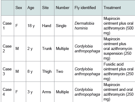 Table 1.Summary of clinical findings, causative larvae, and treatment of the
presented cases.
Table 1.Summary of clinical findings, causative larvae, and treatment of the
presented cases.
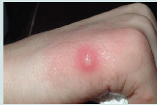 Figure 1a:An erythematous nodule on the ulnar aspect of the right hand,
covered with white petrolatum. This patient had a single painful lesion on
an exposed area, consistent with furuncular myiasis caused by Dermatobia
hominis.c
Figure 1a:An erythematous nodule on the ulnar aspect of the right hand,
covered with white petrolatum. This patient had a single painful lesion on
an exposed area, consistent with furuncular myiasis caused by Dermatobia
hominis.c
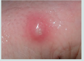 Figure 1b:A close-up view of the central punctum of the nodule, showing the
posterior end of the larva visible through the skin.
Figure 1b:A close-up view of the central punctum of the nodule, showing the
posterior end of the larva visible through the skin.
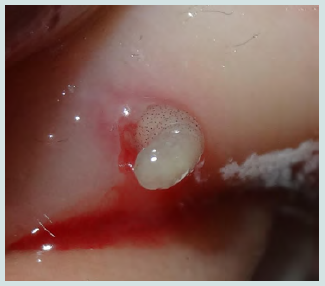 Figure 1c:The D. hominis larva protruding through the skin at its posterior
end. Note the black body spicules.
Figure 1c:The D. hominis larva protruding through the skin at its posterior
end. Note the black body spicules.
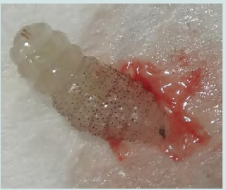 Figure 1d: The D. hominis larva showing characteristic black spicules, which
are absent from the three terminal segments; posterior respiratory spiracles
located away from the skin surface; and mouthparts at the anterior end in
close contact with the skin.
Figure 1d: The D. hominis larva showing characteristic black spicules, which
are absent from the three terminal segments; posterior respiratory spiracles
located away from the skin surface; and mouthparts at the anterior end in
close contact with the skin.
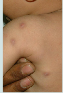 Figure 2a:Multiple nodules on the left side of the upper trunk.
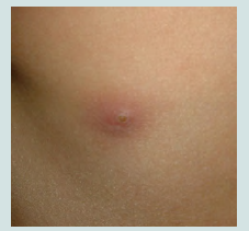 Figure 2b:A close-up view of the lower nodule shown in Figure 2a, with the
larva visible through the skin.
Figure 2b:A close-up view of the lower nodule shown in Figure 2a, with the
larva visible through the skin.
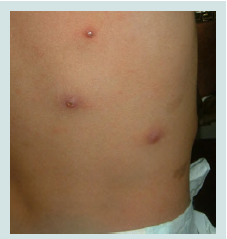 Figure 2c:Three scattered nodules on the right lower back. The involvement
of covered areas and the presence of multiple lesions were consistent with
myiasis caused by Cordylobia anthropophaga.
Figure 2c:Three scattered nodules on the right lower back. The involvement
of covered areas and the presence of multiple lesions were consistent with
myiasis caused by Cordylobia anthropophaga.
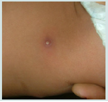 Figure 2d:A lesion on the right lower abdomen before larval extraction.
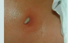 Figure 2e: C.anthropophaga larva on the skin of the right lower abdomen
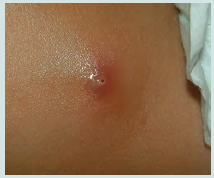 Figure 2f:The lesion shown in Figure 2d after larval removal, with mupirocin
ointment applied.
Figure 2f:The lesion shown in Figure 2d after larval removal, with mupirocin
ointment applied.
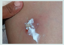 Figure 3a:The medial aspect of the left upper thigh. The larva emerged
spontaneously after application of fusidic acid (Fucidin) ointment to the punctum.
Figure 3a:The medial aspect of the left upper thigh. The larva emerged
spontaneously after application of fusidic acid (Fucidin) ointment to the punctum.
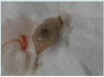 Figure 3b:The extracted C. anthropophaga larva.
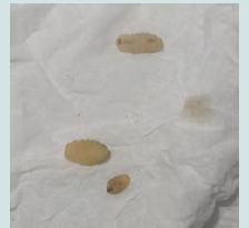 Figure 3c:Larvae self-extracted by the patient and provided to the
investigators.
Figure 3c:Larvae self-extracted by the patient and provided to the
investigators.
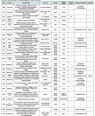 Table 2Reported Cases of Furuncular Myiasis in Arabian Countries.
Case Series
Cutaneous Furuncular Myiasis: A Case Series from Yemen
Alshami MA1*, Alshami AM2, Alshami HM1 and Lutf RM1
1Department of Dermatology, Faculty of Medicine and Medical Sciences,
Sana’a University, Sana’a, Yemen
2Department of Conservative Dentistry, Faculty of Dentistry, Sana’a University, Sana’a, Yemen
2Department of Conservative Dentistry, Faculty of Dentistry, Sana’a University, Sana’a, Yemen
*Address for Correspondence:Mohammad Ali Alshami, Department of Dermatology, Faculty of
Medicine and Medical Sciences, Sana’a University, Sana’a 1064, Yemen. E-mail Id: mohammadalshami62@gmail.com
Submission: 26 July, 2026
Accepted: 24 August, 2026
Published: 28 August, 2026
Copyright: © 2026 Alshami MA, et al. This is an open access
article distributed under the Creative Commons Attribution License,
which permits unrestricted use, distribution, and reproduction in any
medium, provided the original work is properly cited.
Abstract
Myiasis is defined as the infestation of living humans or animals
by the larvae of two-winged flies (order Diptera). The affected sites
may be cutaneous or extracutaneous, with cutaneous myiasis further
classified into furuncular, migratory, and wound forms. Herein, we
present four cases of cutaneous furuncular myiasis that, to the best of
our knowledge, represent the first reported cases of this condition in
Yemen. None of the patients had any history of travel outside Yemen,
making this case series particularly noteworthy. Following clinical
evaluation, an entomologist at our university identified the larvae as
third-instar larvae of Dermatobia hominis in one patient and thirdinstar
larvae of Cordylobia anthropophaga in the remaining three
patients. This case series aims to increase awareness among medical
personnel and to advise individuals at risk of C. anthropophaga
infestation to iron linen and clothing after washing, avoid drying
laundry outside, particularly near the ground and in the shade, and
avoid wearing clothes that have been placed on the ground in the
shade. Additionally, this case series provides evidence that these flies,
despite being native to tropical areas, may be found outside of these
regions owing to international travel, through which eggs or larvae are
inadvertently transported.
Introduction
Myiasis is the infestation of living humans or other vertebrate
animals by the larvae of two-winged flies (order Diptera), which feed,
for at least part of their development, on the living or dead tissues of
the host.[1] Myiasis is classified as cutaneous or extracutaneous, with
cutaneous myiasis further subdivided into the furuncular, migratory,
and wound forms. Among these clinical forms, the furuncular form is
the most common.[2] Although myiasis is endemic in tropical regions
of the Americas and Africa, cases have been reported worldwide.[2]
Nevertheless, a MEDLINE search using the search terms “furuncular
myiasis” and “Yemen” did not identify any previously published
reports of furuncular cutaneous myiasis from Yemen. Therefore, we
report a case series of four patients with this condition.
Case reports
(Table 1) presents a summary of the clinical findings of the four
presented cases.
Case 1
An 18-year-old woman presented with a 3-week history of swelling
on the ulnar aspect of her right hand that had become painful over the
preceding 2 weeks. The patient recalled experiencing an insect bite
at the same site at the onset of symptoms. Cutaneous examination
revealed an erythematous nodule. The posterior end of the larva,
bearing the posterior spiracles, was visible through the central pore
of the nodule. After the area was covered with white petrolatum,
the patient observed slight movement and protrusion of an object
from the center of the lesion. A larva was subsequently removed by
applying gentle pressure. The larva exhibited black circular spicules
on the proximal body segments and two black tubercles at the distal
end. Based on its typical morphology, a third-instar larva of the fly
Dermatobia hominis was suspected and subsequently confirmed by
an entomologist at the Department of Agriculture, Sana’a University
(Figure 1a–1d). Based on the clinical findings (a single lesion on an
exposed area) and the larval morphology, a diagnosis of furuncular
cutaneous myiasis caused by D. hominis was established. The patient
was prescribed topical mupirocin ointment and oral azithromycin
(500 mg), and the lesion showed visible improvement after 1 week.
Case 2:
A 2-year-old boy presented with a 3-week history of multiple
tender nodules on the trunk. The posterior end of the larva, bearing
the posterior spiracles, was visible through the central punctum of
the nodules. A single larva was manually extracted by applying gentle
pressure to the sides of a lesion on the right side of the abdomen and
was subsequently identified by an entomologist as a third-instar larva
of Cordylobia anthropophaga based on its morphological features.
Because the affected areas were normally covered by clothing,
transmission was speculated to have occurred through contact
with damp clothing or via a mechanical carrier, such as a mosquito
(Figure 2a–2f). Topical mupirocin ointment and oral azithromycin
suspension (250 mg) were administered, achieving complete healing
after 1 week.
Case 3:
An 8-year-old boy presented with a 2-week history of two
nodules on the medial aspect of his left thigh. The posterior end of the
larva, bearing the posterior spiracles, was visible through the central
punctum of the nodule. Based on the morphological features of
several larval specimens provided by the patient and a larva extracted
in the clinic, the larvae were identified by an entomologist as third-
instar larvae of C. anthropophaga. As in Case 2, the affected areas
were normally covered by clothing (Figure 3a–3c). Treatment with
topical fusidic acid (Fucidin) ointment and oral azithromycin (250
mg) resulted in complete healing after 10 days.
Case 4:
A 3-year-old girl presented with a 2-week history of multiple
tender nodules on her arms, from which larvae had been manually
extracted by her father, who also provided videos documenting the
extraction process (Videos 1–3). The posterior end of the larva,
bearing the posterior spiracles, was visible through the central
punctum of the nodule. The affected body regions were normally
covered by clothing. The presence of multiple lesions was suggestive
of C. anthropophaga as the causative species. Treatment with topical
mupirocin ointment and oral azithromycin (250 mg) resulted in
complete healing after 7 days.
Discussion
Reporting four cases of furuncular myiasis is very unusual in an
area distant from the natural habitat of myiasis-causing flies, namely
tropical Africa and Central America; however, this unusual finding
may be explained by the possible transport of their eggs or the flies
themselves via transport vehicles such as airplanes, ships, or trains.
Conversely, numerous cases have been reported from countries
distant from the endemic areas of these flies, including Europe, North
America, and Asia. Approximately 12 cases of furuncular myiasis
have been reported in Saudi Arabia, the northern neighbor of Yemen
(Table 2).
In contrast to D. hominis, C. anthropophaga typically penetrates the skin less deeply, although mild pruritus may occur for up to 2 days after infestation. The lesion initially appears as a reddish papule that develops into a boil-like nodule with a central pustule. Erythema may develop in the surrounding tissue within a few days.[7] The mature third-instar larva typically leaves the host after 1–2 weeks, and the posterior spiracles may sometimes be visible through the central pore. Multiple lesions may be associated with systemic symptoms, such as regional lymphadenopathy or malaise.
In contrast to D. hominis, C. anthropophaga typically penetrates the skin less deeply, although mild pruritus may occur for up to 2 days after infestation. The lesion initially appears as a reddish papule that develops into a boil-like nodule with a central pustule. Erythema may develop in the surrounding tissue within a few days.[7] The mature third-instar larva typically leaves the host after 1–2 weeks, and the posterior spiracles may sometimes be visible through the central pore. Multiple lesions may be associated with systemic symptoms, such as regional lymphadenopathy or malaise.
Infestations with C. anthropophaga most commonly occur
during the rainy season. Adult flies oviposit on soiled clothing, which
explains the distribution of lesions on covered body sites, such as the
trunk, buttocks, and thighs, as well as the greater number of lesions
observed in affected individuals. [7] Additionally, the thinner skin
and immature immune system of infants may explain their increased
susceptibility to infestation. In the presented cases the actual route of
infection was unfortunately not established.
In its furuncular form, cutaneous myiasis manifests as boil-like lesions on exposed areas of the skin, such as the face, arms, scalp, or legs, which gradually develop over the course of a few days. A characteristic feature of these lesions is the central punctum, which facilitates air exchange within the lesion and through which the larvae discharge serosanguineous fluid. These puncta can be readily identified with the aid of a magnifying glass or by observing air bubbles that rise when the affected area is submerged in water. The posterior end of the larva, bearing the posterior spiracles, is generally visible through the punctum, as observed in all four cases described above. However, these furuncular lesions resolve rapidly after the larva emerges or is removed.
In its furuncular form, cutaneous myiasis manifests as boil-like lesions on exposed areas of the skin, such as the face, arms, scalp, or legs, which gradually develop over the course of a few days. A characteristic feature of these lesions is the central punctum, which facilitates air exchange within the lesion and through which the larvae discharge serosanguineous fluid. These puncta can be readily identified with the aid of a magnifying glass or by observing air bubbles that rise when the affected area is submerged in water. The posterior end of the larva, bearing the posterior spiracles, is generally visible through the punctum, as observed in all four cases described above. However, these furuncular lesions resolve rapidly after the larva emerges or is removed.
A definitive diagnosis of myiasis is generally established by
extracting and identifying the infesting larva based on its characteristic
morphology. However, in clinical practice, the diagnosis is typically
based on characteristic clinical findings (the site and number of
lesions), particularly in endemic areas. The injection of lidocaine
around the lesion may facilitate painless larval extraction. Treatment
consists of occlusion therapy, manual extraction of the larva, and
topical or oral ivermectin. Topical and, occasionally, systemic
antibiotics are used to prevent secondary bacterial infection and to
facilitate healing, which was the case in the presented cases [2]. This
case series emphasizes the importance of considering furuncular
myiasis in lesions resembling boils, particularly in children living in
resource-limited settings.
Video 1. Case 4. Manual extraction of the larva by the patient’s
father.
Video 2. Case 4. Larva after extraction from the skin.
Video 3. Case 4. Multiple larvae after extraction from the skin.
Video 2. Case 4. Larva after extraction from the skin.
Video 3. Case 4. Multiple larvae after extraction from the skin.
References
Citation
Alshami MA, Alshami AM, Alshami HM, Lutf RM. Cutaneous Furuncular Myiasis: A Case Series from Yemen. J Clin Investigat Dermatol. 2026;14(1): 1