

Journal of Ocular Biology
Download PDF
The two MPS included in this study contain different active disinfecting agents, allowing comparison of their effects. The onestep H2O2 system involves a 3.0% w/v solution of the disinfectant and a dry tablet containing catalase as a neutralizer for H2O2 [25]. While it is common for H2O2 systems to use a platinum disk attached to the lens case for neutralization, catalase neutralization was more practical for the present model. For the purposes of this study, the disinfection step was performed after 1 hour, the time required for the neutralization (and therefore disinfection) of H2O2 to be completed. The PVP-I system includes a dissolving and rinsing solution and a dry tablet with an outer layer containing the primary disinfecting agent and an inner layer containing neutralizing and cleaning agents, as previously described [26-27]. Disinfection was terminated at 30 minutes, ample time for the system to neutralize PVP-I [26-27].
 Table 1.Summary of relevant information on the properties of lens materials tested in the study.
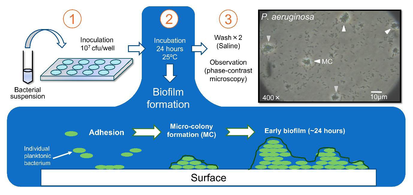 Figure 1:A graphical summary illustrating the process for forming a biofilm with P. aeruginosa within a well plate. (1) Well plates are inoculated at 1 × 10^7 CFU/ well. (2) The plates are incubated for 24 hours at 25°C, and (3) then evaluated for biofilm formation using phase-contrast microscopy. A sequential illustration at the bottom shows the progression from planktonic bacterial adhesion to micro-colony (MC) formation and then early biofilm formation. A representative phase-contrast micrograph of P. aeruginosa micro-colony formation is shown in the top-right panel.
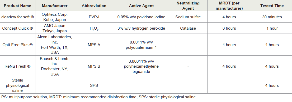 Table 2 A summary of relevant information on the properties of contact lens care solutions tested in the study.
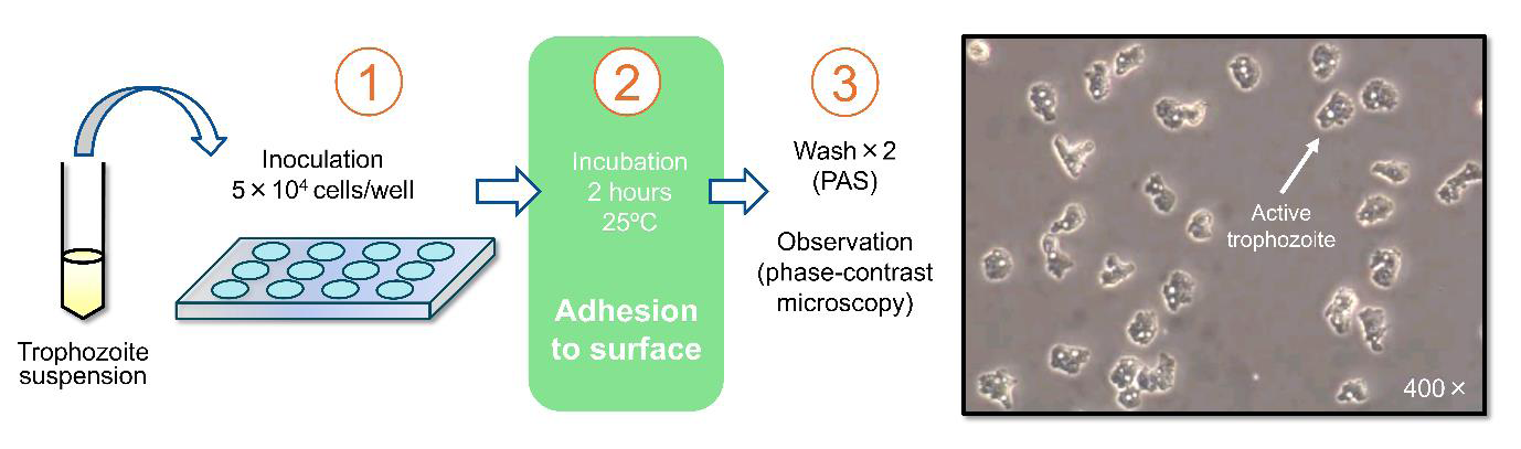 Figure 2:A graphical summary illustrating the process for assessing trophozoite adhesion in a well-plate. (1) An inoculum of (5 × 10^4 cells/well) is added to the wells. (2) Amoebae are allowed 2 hours at 25°C to settle at the bottom of the plate, adhere to the surface, and roam the environment freely. (3) Adherent
trophozoites are then quantified via phase-contrast microscopy. A representative micrograph of active trophozoites post-incubation is shown on the right.
Figure 2:A graphical summary illustrating the process for assessing trophozoite adhesion in a well-plate. (1) An inoculum of (5 × 10^4 cells/well) is added to the wells. (2) Amoebae are allowed 2 hours at 25°C to settle at the bottom of the plate, adhere to the surface, and roam the environment freely. (3) Adherent
trophozoites are then quantified via phase-contrast microscopy. A representative micrograph of active trophozoites post-incubation is shown on the right.
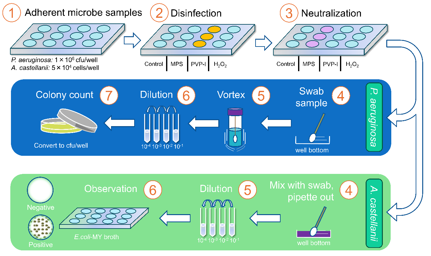 Figure 3: A graphical summary illustrating the method for assessing disinfecting efficacy against adherent microbes. (1) Adherent microbial samples in multi-well plates are created following the process outlined in (Figures 1) and (Figure 2). (2) The sample in the well plate are subjected to either control saline or disinfection treatments including multi-purpose solutions (MPS), povidone-iodine (PVP-I), and hydrogen peroxide (H2O2) for the disinfection times as outlined in (Table 2). (3) This is followed by a standard neutralization step. The recovery and quantification methods differ between the two microbial species, with P. aeruginosa depicted in the top blue panel and A. castellanii depicted in the bottom green panel. Wells containing P. aeruginosa are (4) swabbed, (5) vortexed, (6) serially diluted, and then (7) placed in petri dishes for colony counting. Adherent A. castellanii are (4) dislodged with a swab, and the liquid contents of the well are removed with a pipette. (5) Serial dilutions are performed, then these diluted samples are (6) re-inserted into separate well plates and mixed with E. coli-MY broth, and are later observed to check for positive versus negative activity and proliferation.
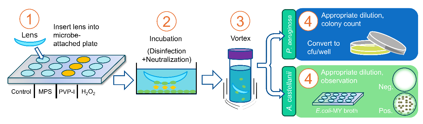 Figure 4:A graphical summary illustrating the method for assessing the impact of care solution on case-lens microbial transmission. (1) A contact lens is placed in a well containing one of the adherent microbial strains, along with one of the care solutions or the saline control. (2) Steps 1-3 from Figure 3 are carried out with the contact lens present in the well. (3) The lenses are removed, inserted into a sealed container containing saline, and vortexed to dislodge transmission red microbes. (4) The recovery and quantification process for each respective microbial species is carried out according to the same method outlined in the blue and green panels of (Figure 3).
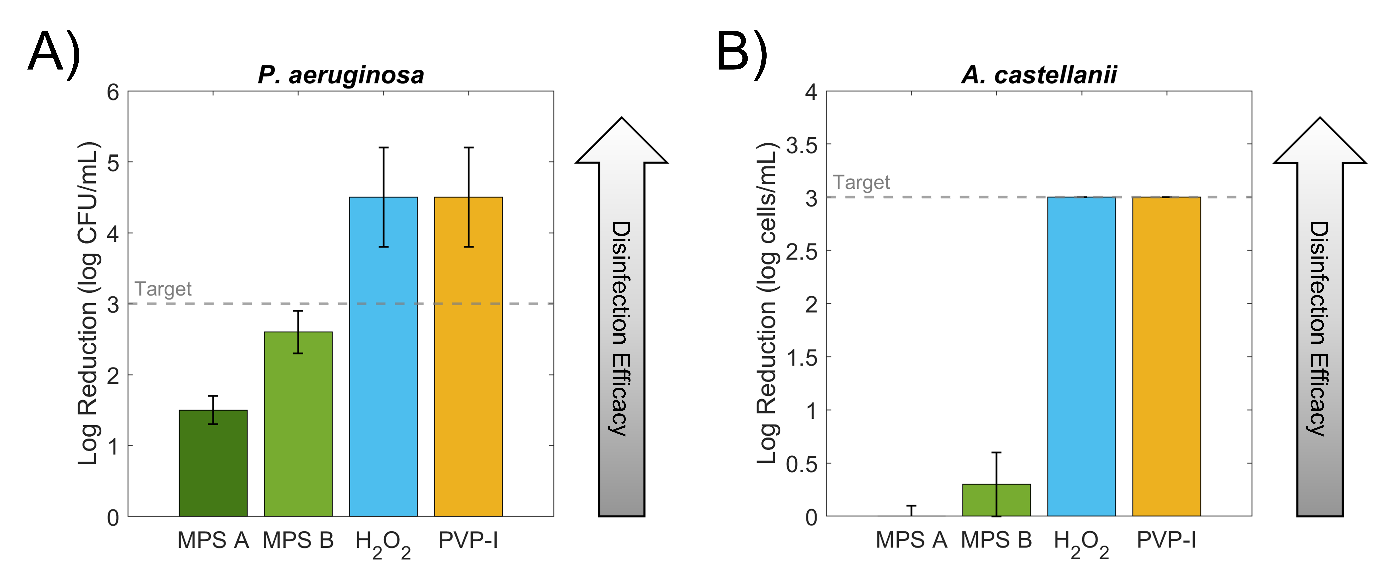 Figure 5:A) A bar graph depicting the comparative disinfection efficacy of two multipurpose solutions (MPS A and MPS B), a 3% hydrogen peroxide solution (H2O2), and a povidone-iodine-based system (PVP-I) against adherent P. aeruginosa. The y-axis represents the log reduction in microbial concentration, with an upward arrow on the right side of the graph indicating increased disinfection efficacy. The target 3-log reduction is indicated by a horizontal dashed line. Error bars represent ±1 standard deviation. B) A bar graph depicting the comparative disinfection efficacy of the aforementioned lens care solutions against adherent A.
castellanii trophozoites. Other graphical indicators are consistent with those of A.
Figure 5:A) A bar graph depicting the comparative disinfection efficacy of two multipurpose solutions (MPS A and MPS B), a 3% hydrogen peroxide solution (H2O2), and a povidone-iodine-based system (PVP-I) against adherent P. aeruginosa. The y-axis represents the log reduction in microbial concentration, with an upward arrow on the right side of the graph indicating increased disinfection efficacy. The target 3-log reduction is indicated by a horizontal dashed line. Error bars represent ±1 standard deviation. B) A bar graph depicting the comparative disinfection efficacy of the aforementioned lens care solutions against adherent A.
castellanii trophozoites. Other graphical indicators are consistent with those of A.
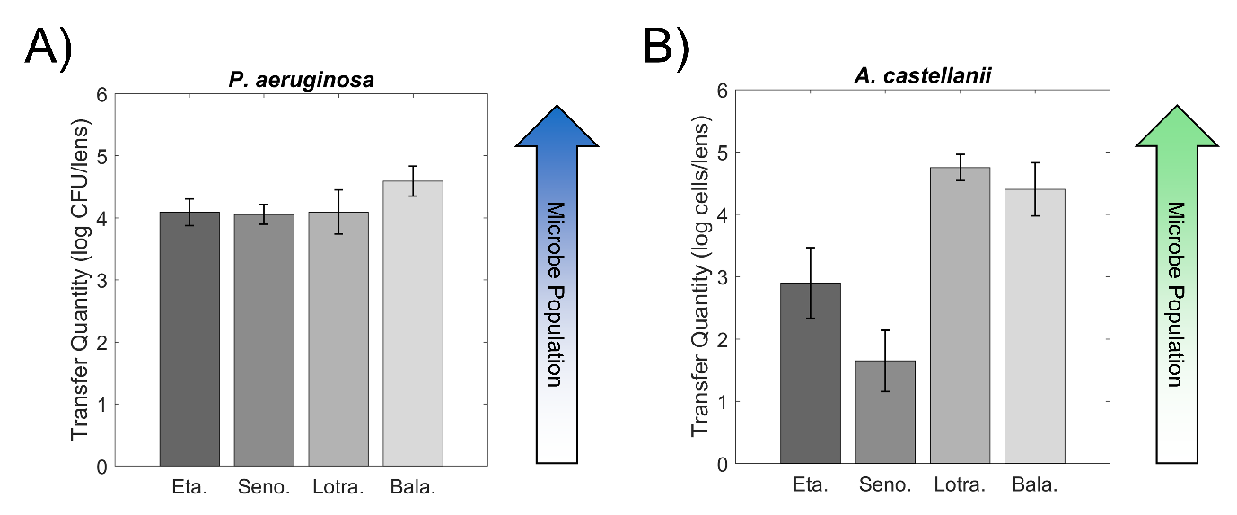 Figure 6:A) A bar graph showing the transmission quantity of P. aeruginosa in log CFU/lens to the four lens materials in saline, and B) a bar graph showing the transmission quantity of A. castellanii trophozoites to each of the four materials in saline. The upward arrows to the right of each graph denote a larger microbial population recovered from the lens. Data are shown as mean ± 1 SD. Lens materials are abbreviated as: etafilcon A (Eta), senofilcon A (Seno), Lotrafilcon B (Lotra), Balafilcon A (Bala).
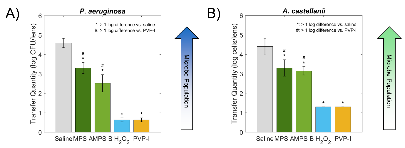 Figure 7:A) A bar graph showing the impact of the two MPS, and the H2O2- and PVP-I-based solutions on the transmission quantity of P. aeruginosa in log CFU/ lens to the Balafilcon A compared to the saline control, and B) a similar bar graph showing the impact of the solutions on the transmission quantity of A. castellanii trophozoites to Balafilcon A. The upward arrows to the right of each graph denote a larger microbial population recovered from the lens. Biologically meaningful differences compared to the control (> 1 log/lens) are indicated by asterisks (*), and meaningful differences compared to PVP-I are indicated by hash symbols (#).
Data are presented as the mean ± 1 SD.
Figure 7:A) A bar graph showing the impact of the two MPS, and the H2O2- and PVP-I-based solutions on the transmission quantity of P. aeruginosa in log CFU/ lens to the Balafilcon A compared to the saline control, and B) a similar bar graph showing the impact of the solutions on the transmission quantity of A. castellanii trophozoites to Balafilcon A. The upward arrows to the right of each graph denote a larger microbial population recovered from the lens. Biologically meaningful differences compared to the control (> 1 log/lens) are indicated by asterisks (*), and meaningful differences compared to PVP-I are indicated by hash symbols (#).
Data are presented as the mean ± 1 SD.
The disinfecting efficacy of the four care systems against adherent A. castellanii is depicted in (Figure 5B). MPS A proved to be ineffective against the protozoan when adhered to a surface, failing to reduce the microbial count (0.0 ± 0.1 log cells/mL) by any significant amount. Likewise, MPS B reduced the microbial count by only 0.3 ± 0.3 log cells/mL, failing to kill trophozoites in a significant proportion. The PVP-I- and H2O2-based systems reduced the microbial count by 3.0 ± 0.0 log cells/mL, achieving a disinfection below the detectable limit.
The transmission of adherent A. castellanii trophozoites to Balafilcon A under the influence of each care solution is depicted in (Figure 7B). 3.3 ± 0.4 log cells/lens of trophozoites, 1.1 log cells/lens less than the saline control, were recovered after exposure to MPS A, an average inhibition of 92.1% (range: 69.1 – 98.0%). Exposure to MPS B resulted in 3.2 ± 0.2 log/lens of recoverable trophozoites, 1.2 log/lens less than the control, and only 0.1 log cell/lens less than MPS A. Proportionally, MPS B inhibited amoebic transmission by 94.4% (range: 83.4 – 98.1%). 1.3 ± 0.0 log cells/lens were recovered from the lenses treated with both PVP-I and H2O2, 3.1 log cells/lens less than the control, and greater than 1 log cells/lens less than either MPS. Proportionally, these solutions inhibited amoebic transmission by 99.9% (range: 99.8 – 100.0%).
Research Article
Quantifying Case-Contact Lens Microbial Transmission and the Influence of Care Solutions
Bahr C*, Takeichi R and Yamasaki K
Kobe Laboratory, Ophtecs Corporation, Kobe, Hyogo, Japan
*Address for Correspondence:Charles Bahr, Kobe Laboratory, Ophtecs Corporation, Kobe, Hyogo, Japan.5-2-4 Minatojima-Minami-Machi Kobe, Hyogo, Japan. Email Id:charlie.bahr@gmail.com
Submission: 03 July, 2026
Accepted: 08 August, 2026
Published: 10 August, 2026
Copyright: © 2026 Bahr C, et al. This is an open access article
distributed under the Creative Commons Attribution License, which
permits unrestricted use, distribution, and reproduction in any medium,
provided the original work is properly cited.
Keywords:Contact Lens; Contact Lens Solution; Contact Lens
Case; Microbial Adhesion, Pseudomonas Aeruginosa; Acanthamoeba
Castellanii; Disinfection
Abstract
Purpose: To quantify the case-to-lens transmission of Pseudomonas
aeruginosa and Acanthamoeba castellanii to soft contact lens
materials and to evaluate the disinfecting efficacy of four common
care systems and their impact using a well-plate model.
Methods: Microbe samples were cultured and allowed to adhere to multi-well plates. The disinfection efficacy of four lens care systems — two multipurpose solutions, a hydrogen peroxide system, and a povidone iodine system – was tested and compared to a saline control. Microbial transmission in saline was quantified by placing one of four lens materials (etafilcon A, senofilcon A, Lotrafilcon B, and Balafilcon A) into contaminated wells and exposing them for 4 hours. The lenses were removed, and their acquired microbial load was quantified. The effect of care systems on transmission was evaluated by disinfecting test lenses in contaminated wells with each product, then quantifying their surviving microbial load. Microbial counts were measured with colony-forming units (bacteria) and the Spearman- Karber method (amoebae).
Results: The povidone iodine and hydrogen peroxide systems achieved > 3-log reductions in both pathogens. Both multipurpose solutions failed to meet the 3-log standard against adherent P. aeruginosa and showed negligible activity against A. castellanii. Bacterial transmission to lenses in saline ranged from 1.1% to 3.9%, with Balafilcon A showing the highest uptake. Amoebic transmission varied by material: Lotrafilcon B (11.3%) and Balafilcon A (5.0%) showed higher affinity compared to etafilcon A and senofilcon A (<0.2%). All care systems significantly inhibited transmission relative to saline (by >1 log/mL), although povidone iodine and hydrogen peroxide were the most effective.
Conclusions: Microbial transmission is dependent on the pathogen and the lens material. Povidone iodine and hydrogen peroxide provide superior protection against transmission compared with multipurpose solutions. This transmission model can be used to evaluate the risk of microbial lens colonization in future work.
Methods: Microbe samples were cultured and allowed to adhere to multi-well plates. The disinfection efficacy of four lens care systems — two multipurpose solutions, a hydrogen peroxide system, and a povidone iodine system – was tested and compared to a saline control. Microbial transmission in saline was quantified by placing one of four lens materials (etafilcon A, senofilcon A, Lotrafilcon B, and Balafilcon A) into contaminated wells and exposing them for 4 hours. The lenses were removed, and their acquired microbial load was quantified. The effect of care systems on transmission was evaluated by disinfecting test lenses in contaminated wells with each product, then quantifying their surviving microbial load. Microbial counts were measured with colony-forming units (bacteria) and the Spearman- Karber method (amoebae).
Results: The povidone iodine and hydrogen peroxide systems achieved > 3-log reductions in both pathogens. Both multipurpose solutions failed to meet the 3-log standard against adherent P. aeruginosa and showed negligible activity against A. castellanii. Bacterial transmission to lenses in saline ranged from 1.1% to 3.9%, with Balafilcon A showing the highest uptake. Amoebic transmission varied by material: Lotrafilcon B (11.3%) and Balafilcon A (5.0%) showed higher affinity compared to etafilcon A and senofilcon A (<0.2%). All care systems significantly inhibited transmission relative to saline (by >1 log/mL), although povidone iodine and hydrogen peroxide were the most effective.
Conclusions: Microbial transmission is dependent on the pathogen and the lens material. Povidone iodine and hydrogen peroxide provide superior protection against transmission compared with multipurpose solutions. This transmission model can be used to evaluate the risk of microbial lens colonization in future work.
Introduction
Contact lens wear poses several inherent risks to ocular health,
the most serious of which is contact lens-related microbial keratitis
[1]. Microbial keratitis (MK) garners much attention from eye care
practitioners due to its morbidity and high risk for permanent corneal
damage and vision loss [1]. Although MK itself is considered a rare
condition, its incidence among contact lens wearers, especially those
who wear reusable or extended-wear soft lenses, is significantly higher
than that among non-wearers [1-3]. Contact lenses can act as a vehicle
for pathogenic organisms, allowing them to remain in prolonged
contact with the ocular surface and providing an opportunity to
invade corneal tissues [4]. One of the major sources of contamination
for reusable lenses is their storage cases. Unhygienic use of contact
lens cases is a known risk factor for MK, and the microbial pathogens
cultured from infected corneas often match those found inside the
wearer’s lens case [2,5].
Two species of interest in MK studies are the bacterium
Pseudomonas aeruginosa and the protozoan Acanthamoeba
castellanii [4,6]. P. aeruginosa is the most common offending
pathogen in contact lens-related MK, accounting for approximately
50% of incidences [7-8]. It is known to form a biofilm – a protective
extracellular matrix composed of polysaccharides, proteins, lipids,
and other biomolecules – when adhered to surfaces such as contact
lens cases [9-11]. Biofilm formation has been shown to increase
bacterial survival during disinfection with contact lens solutions [12-
14]. A. castellanii is a ubiquitous, water-dwelling species of amoeba
that is known to adhere to contact lenses and storage cases [6]. MK
caused by A. castellanii, also known as Acanthamoeba keratitis, is
almost exclusively associated with contact lens wear [6]. Additionally,
up to 20% of contact lens cases have been reported to yield traces of A.
castellanii DNA [15]. Although lens storage cases are a major source
of pathogenic microbes, only a few studies have sought to characterize
and quantify the transmission of case-adherent microbes to contact
lenses [16-17]. Furthermore, to the authors’ knowledge, no studies
have quantified case-to-lens transmission for adherent A. castellanii.
Given that a viable pathogen load in a lens case requires the lens itself
to reach the ocular surface, this presents a notable gap in the literature
regarding the risks of MK. Similarly, a scarcity of data on the capacity
of contact lens care systems to prevent microbial transmission to
lenses, particularly for systems based on oxidative disinfectants such
as hydrogen peroxide (H2O2) and povidone-iodine (PVP-I), warrants
scientific inquiry.
Thus, this study aims to (1) quantify the case-to-lens microbial
transmission of adherent P. aeruginosa and A. castellanii on four
types of reusable soft contact lenses using a novel, simple in-vitro
model, and (2) examine the influence that four different contact lens
care systems – a PVP-I-based system, an H2O2-based system, and two
multipurpose solutions (MPS) – have on this model.
METHODS
Tested Microbial Strains:
A standard strain of Pseudomonas aeruginosa (ATCC 9027) as
specified by ISO 14729:2001 was tested [18]. Acanthamoeba castellanii
(ATCC 50514), an established strain used in disinfection efficacy
tests, was used [19-22]. All culture preparations, inoculations, and
serial dilutions were performed under aseptic conditions in a class II
biosafety cabinet (SCV-1307EC II AB3, Hitachi, Tokyo, Japan).
Tested Lens Materials:
Four different reusable soft contact lens materials were tested:
etafilcon A, senofilcon A, Lotrafilcon B, and Balafilcon A. Relevant
details on the properties of these materials are summarized in (Table
1). Etafilcon A is widely used as a model material to represent the
hydrogel class [23]. Senofilcon A, Lotrafilcon B, and Balafilcon A
likewise each have long tenure in the contact lens market and are
widely used in disinfection testing to represent the SiHy class of
materials [24]. All lenses tested were -3.00 D sphere.
Tested Care Systems:
Four commercially available soft contact lens care systems
were tested: two MPS systems, one H2O2-based system, and one
PVP-I-based system. Relevant details on each of these systems
are summarized in (Table 2). All products were used before their
expiration date.The two MPS included in this study contain different active disinfecting agents, allowing comparison of their effects. The onestep H2O2 system involves a 3.0% w/v solution of the disinfectant and a dry tablet containing catalase as a neutralizer for H2O2 [25]. While it is common for H2O2 systems to use a platinum disk attached to the lens case for neutralization, catalase neutralization was more practical for the present model. For the purposes of this study, the disinfection step was performed after 1 hour, the time required for the neutralization (and therefore disinfection) of H2O2 to be completed. The PVP-I system includes a dissolving and rinsing solution and a dry tablet with an outer layer containing the primary disinfecting agent and an inner layer containing neutralizing and cleaning agents, as previously described [26-27]. Disinfection was terminated at 30 minutes, ample time for the system to neutralize PVP-I [26-27].
Culturing and Scaling Adherent Pseudomonas aeruginosa:
P. aeruginosa (ATCC 9027) was cultured and prepared
according to previously established methods [28]. Briefly, bacteria
were inoculated into Y medium (0.2% Bacto™ tryptone, 0.1% NaCl,
0.2% Bacto™ yeast extract, 0.025% KH₂PO₄, 0.025% K₂HPO₄, 0.01%
(CH₃COO)₂Ca, 0.01% (CH₃COO)₂Mg: pH 7.0) and incubated in a
mechanical shaker at 30°C and 130 rpm for 18 hours. The samples
were harvested, centrifuged (3,000 rpm for 10 min), and the cultured
cells were resuspended in sterile physiological saline (SPS), adjusting
the inoculum to 1.0 × 10^7 colony-forming units (CFU) per mL.
For this study, contact lens cases were substituted for sterile multiwell
plates to ensure consistent conditions for microbial adherence.
We added 1 mL of the bacterial suspension to a multi-well plate and
incubated it at 25°C for 24 hours to allow the bacteria to form an early
biofilm. We removed the supernatant from the wells and rinsed them
twice with SPS to remove any remaining planktonic bacteria. This
generated 1.0 × 10^6 CFU/mL of bacteria adhered to the well plate, a
typical inoculum concentration used in disinfection testing according
to ISO 14729:2001 guidelines [19]. A visual diagram of this process is
depicted in (Figure 1).
Culturing and Scaling Adherent Acanthamoeba castellanii trophozoites:
A. castellanii trophozoites were cultured using a protocol
comparable to established models of Acanthamoeba adhesion
[29-30]. Briefly, we placed ATCC 50514 trophozoites in peptoneyeast-
glucose (PYG) medium for 2 days [31]. After incubation, the
trophozoites were harvested, centrifuged (1,000 × g for 12 min), and
resuspended in Page’s amoeba saline (PAS) to a final concentration of
5.0 × 10^4 cells/mL [32]. This inoculum concentration is consistent
with the recommendations for disinfection testing according to ISO
19045-2:2024 [33]. As mentioned in Section 2.4 for P. aeruginosa,
contact lens cases were substituted for sterile multi-well plates to
ensure consistent conditions for amoebic adherence. 1 mL of the
amoeba suspension was added to each well of a multi-well plate, and
the cells were incubated at 25°C for 2 hours. The supernatant was
removed, the well bottoms were washed twice with PAS to eliminate
non-adherent trophozoites, and the remaining cells were examined
under a phase-contrast microscope to confirm their adhesion. A
visual diagram of this process is depicted in (Figure 2).
Evaluating Disinfection Efficacy of Care Solutions Against Adherent Microbes:
A diagram illustrating the evaluation of disinfection efficacy
against the microbial strains is shown in (Figure 3). For the
disinfection and neutralization steps, the same process was used
for both P. aeruginosa and A. castellanii. The wells containing the
adherent microbes were filled with 4 mL of each test solution (along
with the respective neutralizing tablets provided with the PVP-I
and H2O2 systems) listed in Table 2, and incubated at 25°C for the
corresponding disinfection time. For the two MPS, after removing
the solutions from the well with a pipette, 1 mL of Dey-Engley (D/E)
was added to the neutralizing broth, and the plate was left to stand
for 10 minutes to allow the disinfectants to neutralize [34]. For P.
aeruginosa samples, the neutralizing solution was removed via
pipette, and the adherent bacteria were thoroughly swabbed from
the bottom of each well. The swabbed samples were vortexed for 1
minute in 10 mL of Dulbecco’s phosphate-buffered saline, then serial
dilutions were performed to create triplicate samples at 10^-1, 10^-
2, 10^-3, and 10^-4 of the original concentration [35]. 1 mL of the
diluted samples was mixed with 20 mL of soybean casein digest agar
in petri dishes and incubated at 35°C for 5 days. After incubation,
viable cell counts were measured in colony-forming units and the log
reduction value per well was calculated. Triplicate samples were used
for each count, and each experiment was repeated three times.
For A. castellanii samples, the contents of each well were swabbed
to detach the amoeba from the surface. We then prepared serial
dilutions in PAS to create triplicate samples at 10^-1, 10^-2, 10^-
3, and 10^-4 of the original concentration. We mixed 100 μL of
these samples into a separate well plate containing 1.5 mL of culture
medium per well (Escherichia coli, NBRC 3972, prepared in 500 mL
of malt yeast liquid medium, incubated at 30°C for 18 hours at 130
rpm). After incubating the amoebae at 25°C for 14 days, we observed
the wells under brightfield microscopy, assessed each well for the
presence or absence of active trophozoites, and calculated the log
reduction value using the Spearman-Karber method [36].
Quantifying Microbial Transmission to Contact Lenses:
A visual diagram of the model used to quantify microbial
transmission to contact lenses is shown in Figure 4. To quantify the
transmission of P. aeruginosa and A. castellanii to each contact lens
listed in (Table 1) without the influence of care solutions, the lenses
were first removed from their blister packs and immersed in SPS for
30 minutes to remove blister-pack components. Samples of adherent
microbes were prepared in well plates following the same protocol
as described in Sections 2.4 and 2.5. 4 mL of the appropriate saline
solution (SPS for bacteria and PAS for amoebae) was added to each
well, one lens was inserted, and the plates were incubated at 25°C for
4 hours. The lenses were then removed from the wells, rinsed twice
with 4 mL of the appropriate saline solution, and placed into vials
containing 2 mL of saline, then vortexed for 1 minute to dislodge the
adherent microbes. For P. aeruginosa, serial dilutions were prepared
in Dulbecco’s phosphate-buffered saline to the concentrations
described in Section 2.6. The samples were incubated in soybean casein digest agar at 35°C for 5 days, and colony-forming units (CFUs) were counted to represent the number of bacteria transmitted to the lens. For A. castellanii, serial dilutions of the extracted sample were likewise prepared according to the concentrations in PAS, as described in Section 2.6. We incubated, observed, and calculated the log reduction in trophozoites using the same protocol outlined in Section 2.6. A difference in microbial quantity greater than 1 log (CFU for bacteria, cells for amoebae) per lens was considered biologically meaningful when comparing lenses. Each microbial sample was prepared in triplicate, and the experiment was repeated twice.
described in Section 2.6. The samples were incubated in soybean casein digest agar at 35°C for 5 days, and colony-forming units (CFUs) were counted to represent the number of bacteria transmitted to the lens. For A. castellanii, serial dilutions of the extracted sample were likewise prepared according to the concentrations in PAS, as described in Section 2.6. We incubated, observed, and calculated the log reduction in trophozoites using the same protocol outlined in Section 2.6. A difference in microbial quantity greater than 1 log (CFU for bacteria, cells for amoebae) per lens was considered biologically meaningful when comparing lenses. Each microbial sample was prepared in triplicate, and the experiment was repeated twice.
Disinfection efficacy against microbial transmission to Balafilcon A lenses:
The visual diagram in (Figure 4) also depicts the model used to
assess each care solution’s ability to prevent microbial transmission
to Balafilcon A lenses. Microbial samples and lenses were prepared
according to the same protocol outlined in Section 2.7, except that
one of the four care solutions was inserted into each well instead of
the saline control. The lenses were disinfected for the appropriate
time as specified for each solution in (Table 2), then removed from
the wells and transferred to test tubes containing the neutralizing
solutions. Neutralization was allowed to occur for 10 minutes, then
the lenses were rinsed twice with 4 mL of the appropriate saline
solution and vigorously vortexed in 2 mL of saline for 1 minute. The
surviving microbes were rescaled, cultured, and counted using the
protocol described in Section 2.7. A difference in microbial quantity
greater than 1 log (CFU for bacteria, cells for amoebae) per lens was
considered biologically meaningful when comparing lenses. Each
microbial sample was prepared in triplicate, and the experiment was
repeated twice.
Results
Disinfecting Efficacy of Care Systems Against Adherent Microbial Strains:
The disinfecting efficacy of the four care systems we tested against
adherent P. aeruginosa is depicted in Figure 5A. MPS A showed the
lowest efficacy among the tested care systems, reducing bacteria count
by 1.5 ± 0.2 log CFU/lens. MPS B reduced the bacterial count by 2.6 ±
0.3 log/mL. On the other hand, both PVP-I- and H2O2-based systems
reduced the bacterial count by 4.5 ± 0.7 log CFU/lens, achieving a
disinfection below the detectable limit.The disinfecting efficacy of the four care systems against adherent A. castellanii is depicted in (Figure 5B). MPS A proved to be ineffective against the protozoan when adhered to a surface, failing to reduce the microbial count (0.0 ± 0.1 log cells/mL) by any significant amount. Likewise, MPS B reduced the microbial count by only 0.3 ± 0.3 log cells/mL, failing to kill trophozoites in a significant proportion. The PVP-I- and H2O2-based systems reduced the microbial count by 3.0 ± 0.0 log cells/mL, achieving a disinfection below the detectable limit.
Quantification of microbial transmission to contact lens materials:
The quantification of the transmission of P. aeruginosa from the
well plate to each of the four contact lens materials in sterile saline
is depicted in Figure 6A. 4.1 ± 0.2 log CFU/lens of bacteria were
recovered from etafilcon A. In proportional terms, an average of 1.2%
(range: 0.7 – 2.0%) of the starting count of 1 × 10^6 CFU within the
well was transmitted to the lens. 4.1 ± 0.2 log/lens of bacteria were
recovered from senofilcon A, indicating an average transmission of
1.1% (range: 0.8 – 1.6%). From Lotrafilcon B, 4.1 ± 0.4 log CFU/lens
of bacteria were recovered, indicating an average transmission of
1.3% (range: 0.6% – 2.8%). Balafilcon A exhibited the highest bacterial
transmission, 4.6 ± 0.2 log CFU/lens, corresponding to an average of
3.9% (range: 2.2% – 6.8%).
The quantification of the transmission of A. castellanii
trophozoites to each of the four contact lens materials in PAS is
depicted in (Figure 6B). A recoverable cell count of 2.9 ± 0.6 log
cells/lens was derived from etafilcon A. 1.7 ± 0.5 log CFU/lens were
recovered following the 14-day incubation from senofilcon A, the
lowest of all tested materials. The amoebae showed a high affinity for
Lotrafilcon B, from which 4.8 ± 0.1 log cells/lens were recovered, the
highest of all tested materials. This was followed by 4.4 ± 0.4 log/lens
from Balafilcon A. The recovered log/lens cell counts for etafilcon A
and senofilcon A were statistically indistinguishable from each other
but were significantly lower than those of Lotrafilcon B and Balafilcon
A (p = 0.05, p = 0.05). There was no significant difference found
between Lotrafilcon B and Balafilcon A (p = 0.5).
Impact of care solution on microbial transmission to Balafilcon A:
The impact of each care solution on the transmission of adherent
P. aeruginosa to Balafilcon A is depicted in Figure 7A. 3.3 ± 0.3 log/
lens of bacteria were recovered after exposure to MPS A, more than
1 log CFU/lens less than the saline control. Expressed proportionally,
MPS A inhibited bacterial transmission by an average of 94.9% (range:
6.93 – 18.24%) Exposure to MPS B resulted in 2.5 ± 0.4 log CFU/lens
of recoverable bacteria, 2.0 log CFU/lens less than the saline control,
but only 0.8 log CFU/lens less than MPS A. Proportionally, MBS B
inhibited bacterial transmission by 99.2% (range: 97.3 – 99.7%).
Only 0.6 ± 0.1 log CFU/lens were recovered from lenses treated
with PVP-I and H2O2, 4.0 log/lens less than the saline control, with
a proportional 99.99% inhibition (range: 99.98 – 99.99%). These
solutions significantly inhibited bacterial transmission compared to
both MPS, with a difference greater than 1 log CFU/mL for both.The transmission of adherent A. castellanii trophozoites to Balafilcon A under the influence of each care solution is depicted in (Figure 7B). 3.3 ± 0.4 log cells/lens of trophozoites, 1.1 log cells/lens less than the saline control, were recovered after exposure to MPS A, an average inhibition of 92.1% (range: 69.1 – 98.0%). Exposure to MPS B resulted in 3.2 ± 0.2 log/lens of recoverable trophozoites, 1.2 log/lens less than the control, and only 0.1 log cell/lens less than MPS A. Proportionally, MPS B inhibited amoebic transmission by 94.4% (range: 83.4 – 98.1%). 1.3 ± 0.0 log cells/lens were recovered from the lenses treated with both PVP-I and H2O2, 3.1 log cells/lens less than the control, and greater than 1 log cells/lens less than either MPS. Proportionally, these solutions inhibited amoebic transmission by 99.9% (range: 99.8 – 100.0%).
Discussion
This report serves as a pilot study evaluating the utility of a simple
model for case-to-lens microbial transmission. This model assessed
the quantities of case-adherent P. aeruginosa and A. castellanii
that can be transmitted to reusable contact lenses during storage,
filling an important gap in the literature on contact lens care and
the behavior of these potentially sight-damaging pathogens. It also
provides controlled conditions for a fair, repeatable evaluation of the
performance of contact lens care solutions against case-adherent P.
aeruginosa and A. castellanii.
ISO 14729:2001 provides guidelines for testing the disinfectant efficacy of contact lens solutions against five standard strains: three bacteria and two fungi [19]. These guidelines establish a standard 3-log reduction of planktonic (free-floating) bacteria as the minimum necessary for safe use. However, these guidelines fail to specify whether the same level of disinfection applies to adherent bacteria that have formed a biofilm. Recently, ISO 19045-2:2024 outlined a standardized methodology for testing contact lens solutions against A. castellanii trophozoites and cysts; however, these guidelines do not provide a target log reduction value [36]. For the purposes of this study, a 3-log reduction was considered a logical target reduction for both biofilm bacteria and adherent trophozoites. Among the solutions we tested, only the H2O2- and PVP-I-based systems met the disinfection target for both microbial species.
This model demonstrated disparities in microbial transmission between lens materials for both microbial species tested in the present study, although to varying degrees. Balafilcon A showed a statistically significantly higher affinity for bacteria than the other three lens materials; however, an increase of less than 0.5 log CFU/ lens may not be clinically meaningful. These data suggest that over 4 hours, 1-4% of the original bacterial load in a lens case is a reasonable estimate of the average proportion of adherent P. aeruginosa that can be transmitted from a lens case to a lens in a saline environment. It has been hypothesized that bacterial adhesion may be influenced by a variety of lens characteristics, such as the base material, surface treatments, and water content [4, 37]. These vitro findings suggest that, at least for P. aeruginosa, the effect of the lens material itself may be relatively small compared with other potential factors such as lens modality, case replacement frequency, surface deposits, and patient compliance.
In contrast, transmission of A. castellanii trophozoites was highly dependent on the lens material. Lotrafilcon B and Balafilcon A are high-modulus silicone hydrogel materials that both possess plasma-treated surfaces to enhance wettability. An overall 2-3 log (100-1000×) increase in amoebic uptake demonstrated by Lotrafilcon B and Balafilcon A relative to the other two materials suggests that amoebae may have a particular affinity for these lenses. To the authors’ knowledge, this study is the first to attempt to quantify the case-to lens transmission of Acanthamoeba trophozoites to contact lenses. Given that amoebae must first colonize a contact lens to invade the cornea in Acanthamoeba keratitis, understanding which lenses they have an affinity for will help eye care practitioners make informed decisions when prescribing lenses to their patients.
ISO 14729:2001 provides guidelines for testing the disinfectant efficacy of contact lens solutions against five standard strains: three bacteria and two fungi [19]. These guidelines establish a standard 3-log reduction of planktonic (free-floating) bacteria as the minimum necessary for safe use. However, these guidelines fail to specify whether the same level of disinfection applies to adherent bacteria that have formed a biofilm. Recently, ISO 19045-2:2024 outlined a standardized methodology for testing contact lens solutions against A. castellanii trophozoites and cysts; however, these guidelines do not provide a target log reduction value [36]. For the purposes of this study, a 3-log reduction was considered a logical target reduction for both biofilm bacteria and adherent trophozoites. Among the solutions we tested, only the H2O2- and PVP-I-based systems met the disinfection target for both microbial species.
This model demonstrated disparities in microbial transmission between lens materials for both microbial species tested in the present study, although to varying degrees. Balafilcon A showed a statistically significantly higher affinity for bacteria than the other three lens materials; however, an increase of less than 0.5 log CFU/ lens may not be clinically meaningful. These data suggest that over 4 hours, 1-4% of the original bacterial load in a lens case is a reasonable estimate of the average proportion of adherent P. aeruginosa that can be transmitted from a lens case to a lens in a saline environment. It has been hypothesized that bacterial adhesion may be influenced by a variety of lens characteristics, such as the base material, surface treatments, and water content [4, 37]. These vitro findings suggest that, at least for P. aeruginosa, the effect of the lens material itself may be relatively small compared with other potential factors such as lens modality, case replacement frequency, surface deposits, and patient compliance.
In contrast, transmission of A. castellanii trophozoites was highly dependent on the lens material. Lotrafilcon B and Balafilcon A are high-modulus silicone hydrogel materials that both possess plasma-treated surfaces to enhance wettability. An overall 2-3 log (100-1000×) increase in amoebic uptake demonstrated by Lotrafilcon B and Balafilcon A relative to the other two materials suggests that amoebae may have a particular affinity for these lenses. To the authors’ knowledge, this study is the first to attempt to quantify the case-to lens transmission of Acanthamoeba trophozoites to contact lenses. Given that amoebae must first colonize a contact lens to invade the cornea in Acanthamoeba keratitis, understanding which lenses they have an affinity for will help eye care practitioners make informed decisions when prescribing lenses to their patients.
It is important to acknowledge that while proportions of bacterial
transmission are readily calculable using the methods employed
in this study, the sensitivity of the Spearman-Karber method and
the 14-day incubation period complicate these projections for
Acanthamoeba. Notably, the recovery values in log cells/lens from
Lotrafilcon B and Balafilcon A in saline were highly similar to the
initial inoculum size (5.0 × 10^4 cells/mL, or 4.7 log\mL). Although
it is unlikely for the well’s entire population to move to the lens, it is
clear that Acanthamoeba possesses a strong enough affinity for these
lenses to transmit in sufficient numbers to saturate the assay.
The main purpose of contact lens care systems is to provide
adequate disinfection of lenses and cases to prevent infection during
wear [38]. This study has shown how MPS have low efficacy against
potentially pathogenic Pseudomonas and Acanthamoeba adhered to a
lens case. However, testing lens case disinfection alone is insufficient
- evaluating how care systems impact case-to-lens transmission is
critical to assessing real-world performance, as microbes must first
move from the case to the lens – their vehicle to the eye – before they
can potentially invade the cornea. The model presented in this study
provides consistent conditions for identifying meaningful differences
among care solutions while simulating the realistic route a microbial
load might take to reach the ocular surface. This allows the direct
observation of not only the disinfection efficacy against adherent
microbes in the simulant lens case, but also the impact care solutions
have upon lens-case transmission specifically.
In general, higher disinfection efficacy against case-adherent
microbes reduces microbial transmission from the lens to the case,
as is evidenced by the oxidative disinfectants H2O2 and PVP-I. H2O2
primarily acts by generating reactive oxygen species that initially
damage cell membrane components, gradually working their way
inward to intracellular proteins and DNA. PVP-I, which releases
free iodine in solution, derives its oxidizing power from its inherent
elemental properties as a halogen, quickly penetrates microbial cell
membranes, and begins disrupting intracellular function upon
contact. PVP-I has been shown to be highly effective against bacteria
within biofilms and rapidly tranquilize amoebic activity [13-14,
26, 39]. This report confirms that both oxidative solutions provide
a powerful barrier to lens adherence and were highly effective at
preventing the case-to-lens transmission of adherent P. aeruginosa
and A. castellanii trophozoites.
The MPS tested in this study demonstrated a limited capacity to
prevent transmission compared to oxidative systems. Although the
recovery data from Balafilcon A for P. aeruginosa and A. castellanii
indicate that MPS A and MPS B may somewhat inhibit microbial
motility compared to saline, these solutions still allowed a sizeable
microbial load to survive and colonize the lens. The primary
disinfecting agent in MPS A, polyhexamethylene biguanide (PHMB),
and the primary disinfecting agent in MPS B, polyquaternium-1 (PQ-
1), are large, heavy molecules that disrupt microbial cell membrane
integrity [40-43]. Biofilms can be a robust enough barrier to block
these larger compounds from reaching the bacteria [11-13]. Surfaceadhered
Acanthamoeba trophozoites are also known to be resistant
to MPS disinfectants and can fully regain motility when no longer
exposed [39, 44].
P. aeruginosa and A. castellanii are two clinically pertinent
microbial species; however, more work is needed to explore the
specific interactions of other pathogenic microorganisms with
lens cases, materials, and care solutions. Likewise, an evaluation
of microbial colonization with additional lens materials and care
systems is warranted.
In conclusion, the model presented in this study provides a compendious method for evaluating microbial adhesion, case-to-lens transmission, and disinfection. The transmission of P. aeruginosa was relatively consistent across the four lens materials, while A. castellanii showed a significantly higher affinity for Lotrafilcon B and Balafilcon A compared to etafilcon A and senofilcon A. Care solutions containing oxidative disinfecting agents H2O2 or PVP-I are reliable tools for preventing microbial colonization of both cases and lenses, and should continue to be considered by contact lens wearers. Eye care practitioners should persist in their efforts to educate patients on the limitations of MPS and the potential risks associated with inadequate biocidal efficacy.
In conclusion, the model presented in this study provides a compendious method for evaluating microbial adhesion, case-to-lens transmission, and disinfection. The transmission of P. aeruginosa was relatively consistent across the four lens materials, while A. castellanii showed a significantly higher affinity for Lotrafilcon B and Balafilcon A compared to etafilcon A and senofilcon A. Care solutions containing oxidative disinfecting agents H2O2 or PVP-I are reliable tools for preventing microbial colonization of both cases and lenses, and should continue to be considered by contact lens wearers. Eye care practitioners should persist in their efforts to educate patients on the limitations of MPS and the potential risks associated with inadequate biocidal efficacy.
References
Citation
Bahr C, Takeichi R, Yamasaki K. Quantifying Case-Contact Lens Microbial Transmission and the Influence of Care Solutions. J Ocular Biol. 2026;10(1): 1.